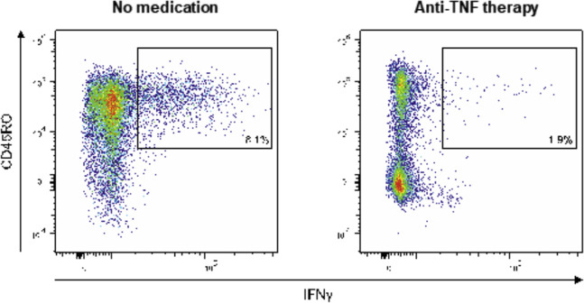
Supplementary Figure 6.
Representative flow cytometry plots of cellular nucleocapsid reactivity. PBMCs were cocultivated with peptide pools covering the N protein for 60 h, restimulated, and analyzed using intracellular cytokine staining. Dot plots show CD4+ and CD8+ T cells that produced IFN-γ in response to stimulation nucleocapsid peptides in a patient with IBD without medication (left) and a patient with anti-TNF therapy (right).

