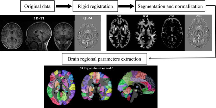
FIGURE 1.

Schematic diagram of the image processing and parameter extraction. CSF, cerebrospinal fluid ; GM, gray matter; QSM, quantitative susceptibility mapping; WM, white matter

Schematic diagram of the image processing and parameter extraction. CSF, cerebrospinal fluid ; GM, gray matter; QSM, quantitative susceptibility mapping; WM, white matter