. 2020 Oct 7;10(61):37168–37181. doi: 10.1039/d0ra06846b

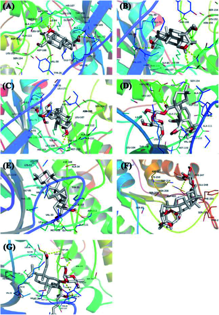
Fig. 7. The 3D docking results of ten core protein molecules and seven core components are visualized. The structure of the compound is represented by a stick, the different branches of the protein are represented by different colors, and the yellow dotted line represents its hydrogen bond, which marks the position of the hydrogen bond and the compound in the compound. (A) Interaction between 3β,23-dihydroxy-1,12-dioxo-olean-28-oic acid and MAPK14; (B) interaction between 3β,23,27-trihydroxy-1-oxo-olean-12-ene-28-oic acid and MAPK14; (C) interaction between 2α,3β,23-trihydroxyurs-11-oxo-12-ene-28-oic acid and MAPK14; (D) interaction between arjunglucoside II and MAPK14; (E) interaction between α-boswellic acid and MAPK14; (F) interaction between 2α,3β,23-trihydroxyoleana-11,13(18)-dien-28-oic acid and CASP3; (G) interaction between cyclocarioside K and MAPK14.

Fig. 7