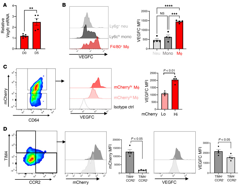
Figure 3. Selective expression of Vegfc in cardiac macrophages after MI.
(A) Sorted cardiac macrophages 5 days after MI were assessed for Vegfc mRNA expression compared with non-MI mice. n = 5/group. **P < 0.005, by unpaired t test. (B) Histogram of VEGFC expression in the indicated cell types by quantitative flow cytometry using cardiac extracts, 7 days after MI. Neutrophils (Neu) were defined as CD11b+Ly6g+; monocytes (Mono) were CD11b+Ly6g–Ly6chiF4/80–; macrophages (Mac) were defined as CD11b+Ly6g–Ly6cloF4/80+CD64+. n = 4 per group. ***P < 0.0002 and ****P < 0.0001, by 2-way ANOVA followed by Tukey’s test. (C) mCherry mice were subjected to coronary ligation to track the uptake of cardiac antigens. Cardiac macrophages with higher levels of mCherry signal also expressed higher levels of VEGFC. n = 4 per group. P < 0.01, by 2-tailed, unpaired t test. (D) Cardiac macrophages were further classified by TIM4 (resident) or CCR2 (recruited) expression. TIM4+ resident macrophages had a higher frequency of mCherry uptake and expressed higher levels of VEGFC. n = 4 per group. Data were pooled from 2–3 independent experiments. P < 0.05, by 2-tailed, unpaired t test. Data are presented as the mean ± SEM.

