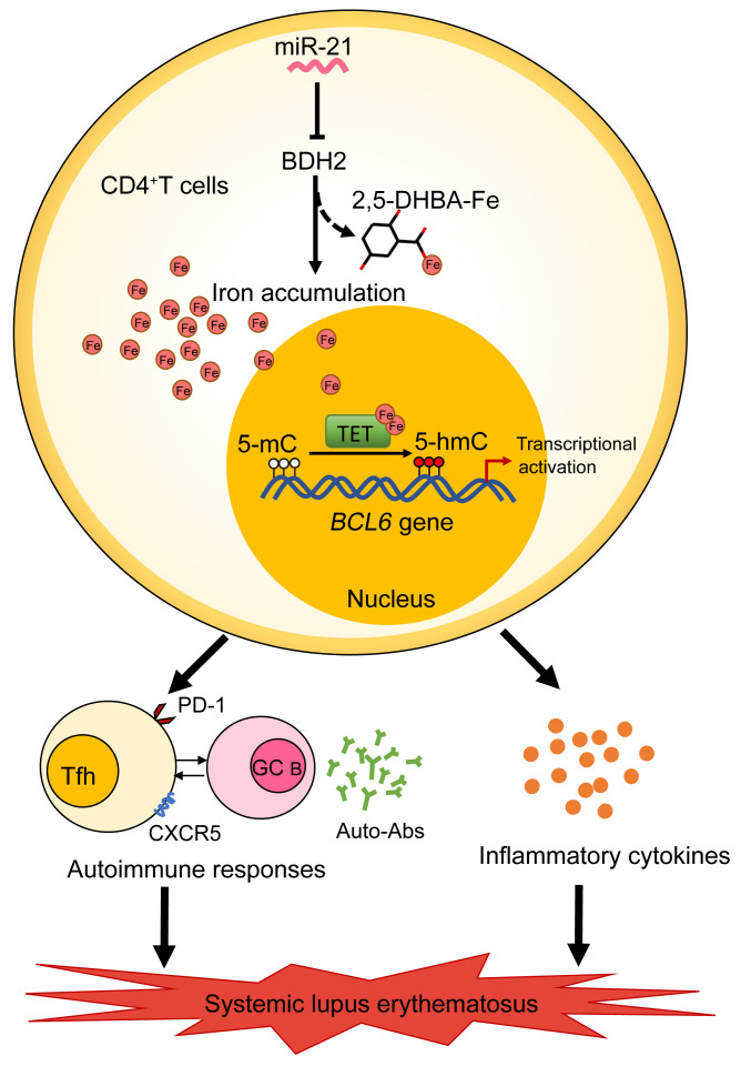
Figure 10. Schematic illustration of the contribution of iron overload to the pathogenic T cell differentiation and pathogenesis of SLE.
In lupus CD4+ T cells, iron accumulation promotes Tfh cell differentiation and Tfh cell–mediated autoimmune responses, autoantibody production, as well as inflammatory cytokine secretion, driving disease progression of lupus. Mechanically, miR-21 represses BDH2 to induce iron accumulation in lupus CD4+ T cells by limiting the synthesis of siderophore 2,5-DHBA, which enhances Fe2+-dependent TET enzyme activity and promotes BCL6 promoter hydroxymethylation and transcription activation, leading to excessive Tfh cell differentiation in SLE. Together, iron overload is an important inducer of the autoimmune response in lupus, and maintaining iron homeostasis will provide a good way for therapy and management of SLE.

