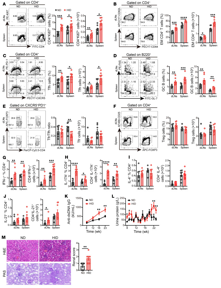
Figure 2. HID contributes to pathogenic T cell differentiation in lupus mice.
3-week-old female MRL/lpr mice were fed with a normal iron diet (ND, 50 mg/kg, n = 8) or a high-iron diet (HID, 500 mg/kg, n = 8) for 20 weeks. (A–F) Representative flow cytometry and (A) quantification of CD4+Ki67+ cells, (B) CD4+CD44+CD62L– effector memory (EM) cells, (C) CD4+CXCR5+PD-1+ Tfh cells, (D) B220+GL-7+FAS+ GC B cells, (E) CD4+CXCR5+PD-1+FOXP3+ Tfr cells, and (F) CD4+CD25+FOXP3+ Tregs in MRL/lpr mice fed with ND or HID. (G–J) Quantification of (G) CD4+IFN-γ+ cells, (H) CD4+IL-17A+ cells, (I) CD4+ IL-4+ cells, and (J) CD4+ IL-21+ cells in MRL/lpr mice fed with ND or HID. (K) Serum levels of anti-dsDNA IgG in MRL/lpr mice fed with ND or HID. (L) Urine protein of MRL/lpr mice fed with ND or HID. (M) Representative morphology (by H&E and PAS staining) and histological scoring of kidneys of MRL/lpr mice after 20 weeks of ND or HID treatment. Scale bar: 50 μm. Cells were isolated from dLNs and spleens of 23-week-old ND- and HID-treated mice. Data are shown as mean ± SEM. Data are representative of 2 independent experiments. *P < 0.05, **P < 0.01, ***P < 0.001, ****P < 0.0001 (unpaired 2-tailed Student’s t test for A–K and unpaired 2-tailed Mann-Whitney U tests for L and M).

