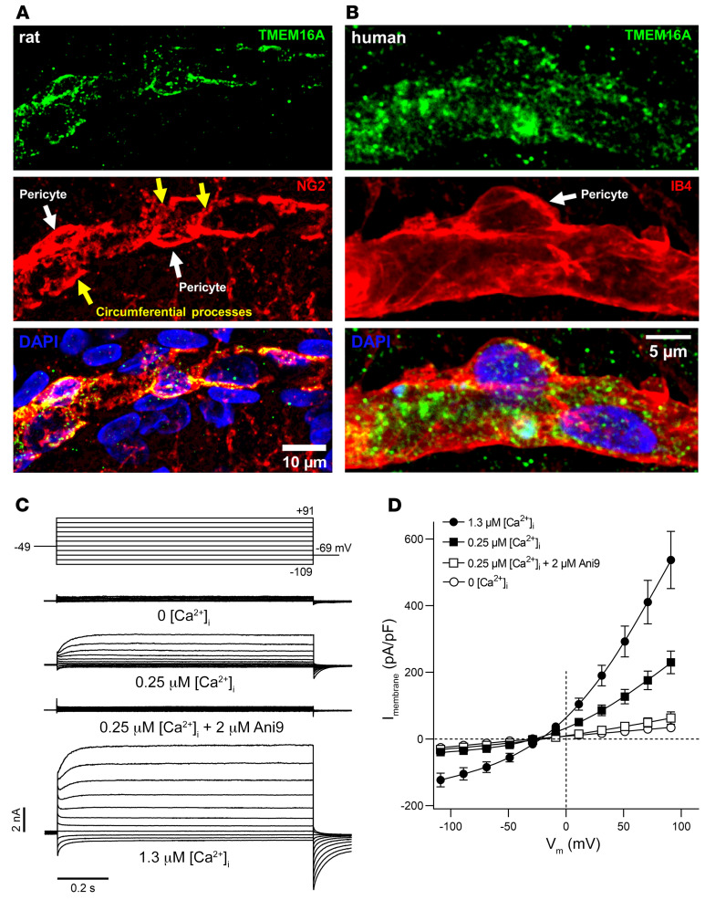
Figure 1. Cortical pericytes express functional TMEM16A channels.
(A) TMEM16A expression in the soma and circumferential processes (yellow arrows) of NG2-labeled pericytes (white arrows) in a representative fixed cortical slice of a P21 rat. Scale bar: 10 μm. (B) TMEM16A expression in a pericyte labeled using isolectin B4 (IB4) in a fixed cortical slice of a 40-year-old human (representative of data from 5 participants). Scale bar: 5 μm. (C) Representative family of whole-cell TMEM16A currents recorded from individual rat cortical pericytes, using pipette solutions designed to isolate Cl– currents and various free [Ca2+]i (nominally 0, 0.25, or 1.3 μM), in the absence or presence of Ani9 (2 μM). The voltage protocol is illustrated at the top, shown after correction for liquid junction potential. (D) Mean whole-cell TMEM16A current density versus voltage relationships in cortical pericytes (n = 9–14), with various [Ca2+]i, in the absence or presence of Ani9.

