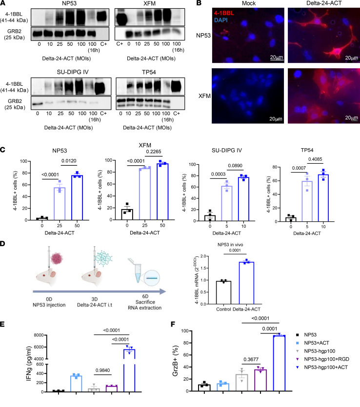
Figure 1. Characterization of Delta-24-ACT functionality in DIPG.
(A) 4-1BBL protein expression in NP53, XFM, SU-DIPG IV, and TP54 cells infected with Delta-24-ACT at the indicated MOIs, as determined by Western blotting. C+, 4-1BBL recombinant protein. (B) Representative immunofluorescence images of 4-1BBL expression in NP53- and XFM-infected cells compared with mock-infected cells. Scale bar: 20 μm. (C) 4-1BBL protein expression in the membranes of murine and human cells infected with Delta-24-ACT at the indicated MOIs, as determined by flow cytometry. The percentage of 4-1BBL+ cells is shown. One-way ANOVA was performed (n = 3, each group), and P values are shown above respective bars. Data are shown as the mean ± SEM. (D) Schedule of the experiment for the in vivo determination of 4-1BBL expression, and evaluation of 4-1BBL protein expression in NP53 tumors from control- or Delta-24-ACT–treated mice (n = 3), as determined by Q-PCR. Data are shown as the mean ± SEM. (E) IFN-γ and (F) granzyme B production by CD8+ lymphocytes. CD8+ T cells from PMEL mice were cocultured with NP53 cells infected with Delta-24-RGD, Delta-24-ACT (MOI = 100), or the mock control. CD8+ lymphocytes activated with CD3 and CD28 but not NP53 cells, and CD8+ lymphocytes activated with CD3, CD28, and 4-1BB antibody were used as negative and positive controls for the experiment, respectively. One-way ANOVA was performed (n = 3, each group), and P values are shown above respective bars. Data are shown as the mean ± SEM.

