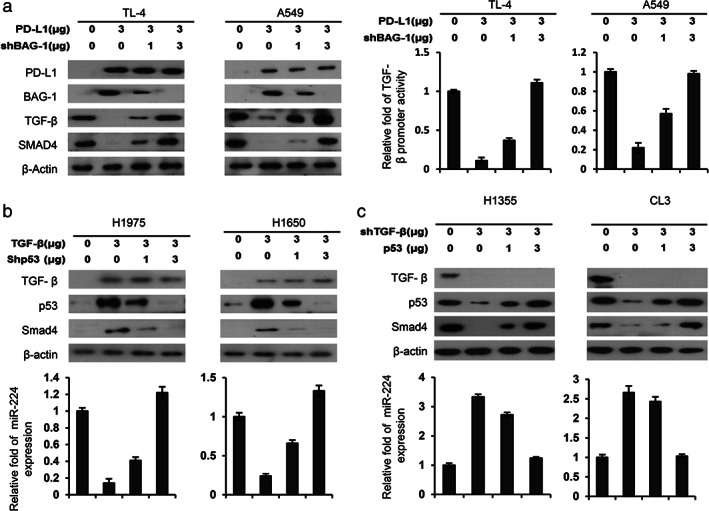
FIGURE 5.

The decrease in TGF‐β1 and SMAD4 expression may be responsible for PD‐L1‐mediated cell invasion in lung cancer cells. (a) TL4 and A549 cells were transfected with PD‐L1 and/or shBAG‐1 for 48 hours and then the expression of PD‐L1, BAG‐1, TGF‐β1, and SMAD4 was evaluated by Western blotting. (b) H1975 and H1650 cells were transfected with TGF‐β1 expression plasmid and/or shp53 and then the expression of TGF‐β1, p53, and SMAD4 was evaluated by Western blotting. (c) H1355 and CL3 cells were transfected with shTGF‐β1 and/or p53 expression plasmid and then the expression of TGF‐β1, p53, and Smad4 was evaluated by Western blotting. The change in miR‐224 levels by ectopic TGF‐β1 expression and/or p53 silencing was evaluated by real‐time PCR. The change in invasion ability of H1975 and H1650 cells was determined by Boyden chamber assays. All experiments were performed independently and in triplicate
