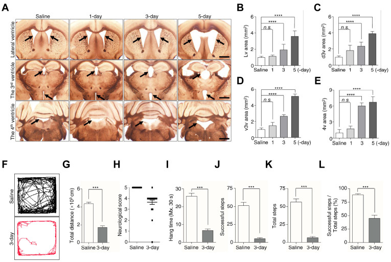
Fig. 1.
Kaolin-induced hydrocephalus mice show ventricular enlargement and motor disturbances. (A) Coronal sections showed ventricles saline and 1, 3, 5-day after kaolin injection. (B-E) Bar plots were showed the Lv, d3v, v3v, and 4v areas. Black arrowheads indicate the ventricles, in the 3v figures, the arrowheads represent the d3v (top) and the v3v (bottom). (F, G) Movement activity was measured for 10 min in the open-field test. (H) The neurological function was scored by a 5-point paradigm and plotted. (I-L) Bar plots showed the results were calculated for 30 s in a horizontal grid test. Bregma in Lv (+0.14 mm), in 3v (−1.12 mm to −1.46 mm), in 4v (−5.88 mm). Ventricles size (n = 6), behavior test (n (saline) = 16, n (3-day) = 24 for the neurological score, n (3-day) = 23 for the open-field test and horizontal grid test, **P < 0.01, ***P < 0.001; n.s., not significant). Scale bar: A: 200 μm.

