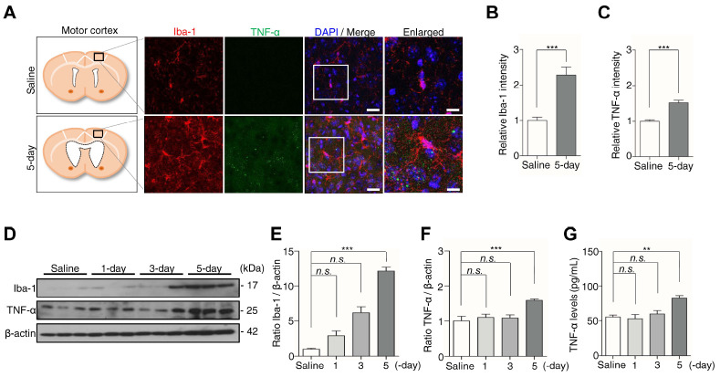
Fig. 2.
Kaolin-induced hydrocephalus mice activate microglia at the 5-day. (A) The motor cortex was stained for Iba-1 (red) and TNF-α (green). (B, C) The immunofluorescence intensity of Iba-1 and TNF-α was quantified. (D) The expression of Iba-1 and TNF-α in the motor cortex was analyzed by western blotting. (E, F) The intensity value of Iba-1 and TNF-α was shown. (G) The TNF-α level was analyzed by ELISA. Western blotting (n = 3, from three independent samples performed twice independently), immunofluorescence and ELISA (n = 6, **P < 0.01, ***P < 0.001; n.s., not significant). Scale bar: A: 20 μm, the enlarged images: 10 μm.

