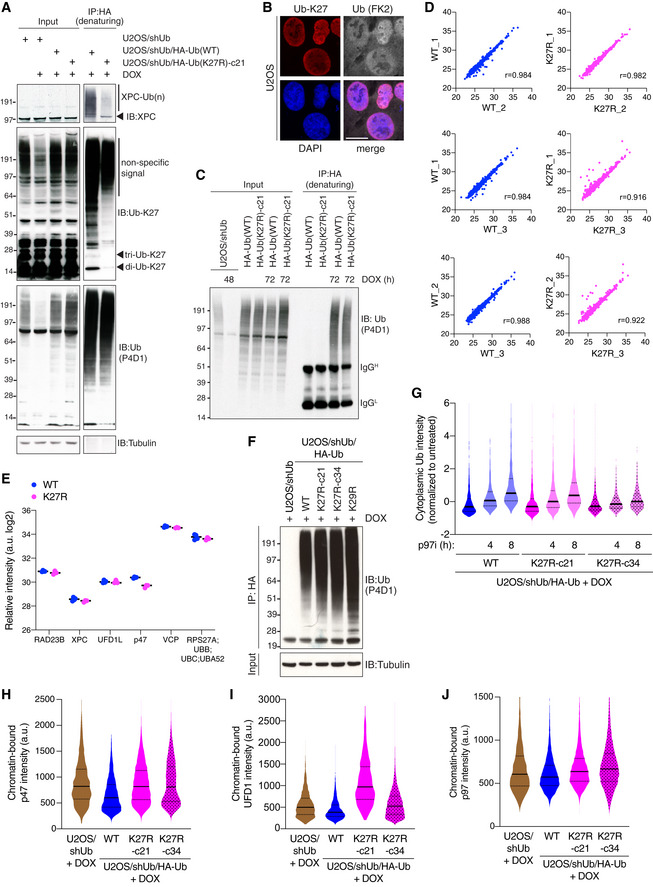
Figure EV3. (related to Fig 3) K27‐linked ubiquitylation predominantly occurs in the nucleus and affects the integrity of the p97 machinery.

-
AU2OS/shUb and DOX‐treated U2OS/shUb/HA‐Ub cell lines were lysed, subjected to HA IP under denaturing conditions and immunoblotted with indicated antibodies.
-
BRepresentative images of U2OS cells co‐immunostained with antibodies to K27‐linked Ub (Ub‐K27) and total Ub conjugates (FK2). Scale bar, 10 μm.
- C
-
DCorrelation analysis comparing label‐free quantitation values of protein groups from individual replicate HA‐Ub conjugate samples isolated as in Fig 3D.
-
ERelative intensity of peptide detection for indicated proteins in U2OS/shUb/HA‐Ub(WT) and U2OS/shUb/HA‐Ub(K27R) whole cell extract analyzed by MS (black bars, median; n = 3 technical replicates). Full proteome data are shown in Dataset EV2.
-
FDOX‐treated U2OS/shUb (48 h) and U2OS/shUb/HA‐Ub (72 h) cell lines were lysed, subjected to HA IP under denaturing conditions and immunoblotted with indicated antibodies as in Fig 3F and G.
-
GDOX‐induced U2OS/shUb/HA‐Ub cell lines treated or not with p97i for the indicated times were fixed and immunostained with Ub conjugate‐specific antibody (FK2). Levels of Ub conjugates in the cytoplasm were quantified using QIBC (solid lines, median; dashed lines, quartiles; > 1,000 cells analyzed per condition).
-
HDOX‐induced U2OS/shUb and U2OS/shUb/HA‐Ub cell lines were pre‐extracted, fixed and immunostained with p47 antibody, and analyzed by QIBC (solid lines, median; dashed lines, quartiles; > 3,000 cells analyzed per condition).
-
IAs in (H), except cells were immunostained with UFD1 antibody.
-
JAs in (H), except cells were immunostained with p97 antibody.
Data information: Data (A,B,F‐J) are representative of three independent experiments with similar outcome.
