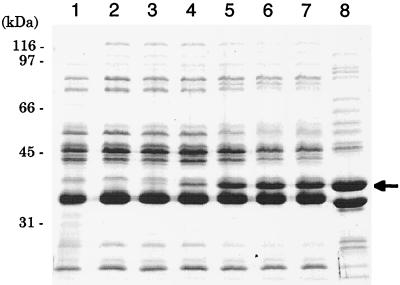
FIG. 2.
Expression of OmpF in the outer membrane of P. aeruginosa and E. coli cells. Cells were grown at 37°C for 18 h on L agar containing various amounts of IPTG. The outer membrane proteins were prepared, and sodium dodecyl sulfate-polyacrylamide gel electrophoresis was performed as described previously (10). Lanes: 1, PAO1 (IPTG, 0 mM); 2, PAO1/pKMF012 (IPTG, 1 mM); 3, PAO1/pKMF010 (IPTG, 0 mM); 4, PAO1/pKMF010 (IPTG, 0.02 mM); 5, PAO1/pKMF010 (IPTG, 0.06 mM); 6, PAO1/pKMF010 (IPTG, 0.25 mM); 7, PAO1/pKMF010 (IPTG, 1.0 mM); 8, E. coli K-12 (IPTG, 0 mM). An arrow shows the position corresponding to E. coli OmpF (37 kDa).

