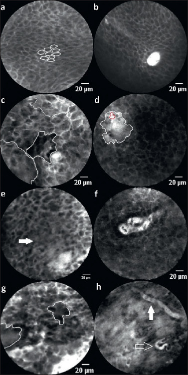
Figure 1.

Description: (a) inconspicuous squamous epithelium with a classic “honeycomb” pattern and (b) capillary loops (rated as -2 points). (c) Inhomogenous global impression with areas of different grey levels. (d) Substantial leakage of fluorescein (white area) around a capillary (red area) indicates malignantly differentiated or altered inflammatory mucosa. (e) The arrow marks elapsed cell borders. Besides, there is vascular leakage (lower right) and an inhomogeneous cell pattern with varying cell sizes. In conclusion, this image is scored as 4 points. (f) Dilated, atypical capillary loop, which is interpreted as suspicious. (g) Cell conglomerates (circled areas), inhomogeneous cell pattern and size adjacent to dilated vessels and leakage (scored 5 points). (h) Atypical blood vessels with a horizontal course in the superficial epithelial layer (white arrow) and a corkscrew-like shape as a sign of neoangiogenesis (transparent arrow). This image is scored as 5 points in total.
