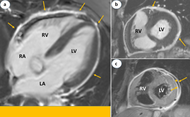
Fig. 2.
Cardiac magnetic resonance images of a 73-year-old male with acute pericarditis. Four-chamber (a) and short axis (b) views showing LGE restricted to the pericardium (yellow arrows). c is STIR image short axis view and demonstrates edema of both in the visceral and parietal layers of the pericardium (yellow arrows) separated by a mild pericardial effusion (asterisk). LA, left atrium; RA, right atrium; LV, left ventricle; RV, right ventricle; LGE, late gadolinium enhancement; STIR, short-tau inversion-recovery.

