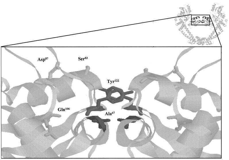
FIG. 1.
QRDR of GyrA. The image at the top right shows a ribbon representation (RasMol) of the 59-kDa N-terminal fragment of GyrA (17). The QRDR is in dark gray. The main image is an expanded version of this region showing the active-site tyrosine (Tyr122) and residues of the QRDR. Ser83 and Asp87 are solvent exposed in this structure and have been mutated to Ala in this study.

