
Figure 1. AMPK is activated following chemotherapy and is a source of sensitivity.
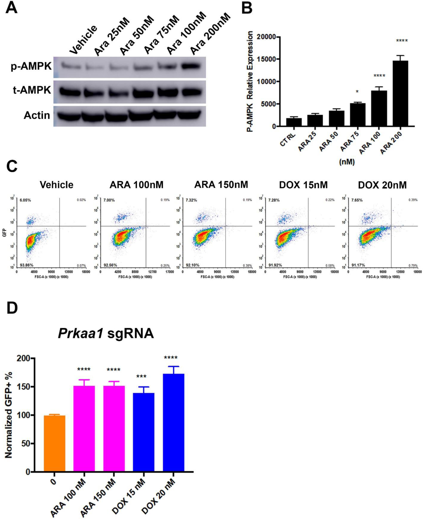
A) Western blot for phosphorylated (p-AMPK) or total (t-AMPK) AMPK alpha subunit. Murine AML cells (MFL2) were treated with the indicated amount of cytarabine (Ara) for 16 hours and lysates blotted as indicated. Actin served as a loading control. B) Densitometry of triplicate Western blots as done in (A) normalized to actin. C) Competition assay. Cas9 expressing Murine AML cells were partially infected with an sgRNA targeting Prkaa1 with a GFP reporter. Cells were treated as indicated for 72 hours and viable fraction analyzed for GFP expression by flow cytometry. Representative contour plots are shown for each treatment tested. D) Competition assay. Normalized GFP+ percentages for each conditioned tested. Shown are the mean values of three independent experiments each done in triplicate. *=p value <0.05, ***=p value <0.005, ****=p value <0.001.