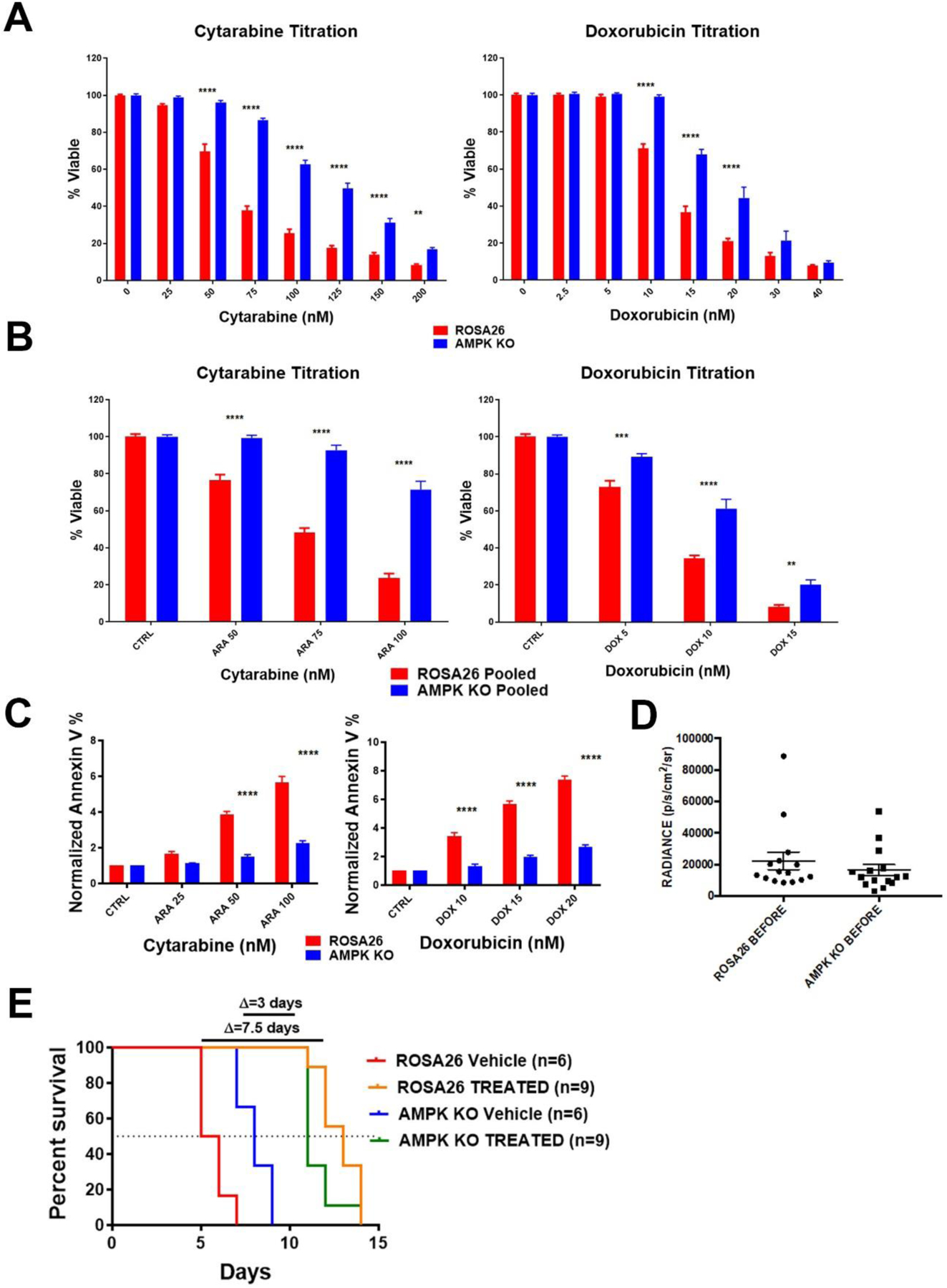
Figure 3. AMPK is a source of sensitivity to chemotherapy in AML.

A) Viability assays. AMPK KO and Rosa26 clonal isolates were exposed to the indicated chemotherapy for 72 hours and viability assessed. B) Pooled clones of AMPK KO or control Rosa26 cells were exposed to the indicated chemotherapy for 72 hours and viability assessed. C) AnnexinV assays. AMPK KO and Rosa26 cells were exposed to the indicated chemotherapy for 72 hours, stained with annexin V and analyzed by flow cytometry. Shown are the means of three independent experiments with the data normalized to each vehicle. D) Quantified bioluminescence. Mice were injected with either AMPK KO or control Rosa26 cells and subjected to bioluminescence imaging. Engraftment at time of treatment initiation is shown. E) Kaplan-Meier curves for vehicle or chemotherapy treated mice. Change in median survival in days is shown above the graph. *=p value <0.05, **=p value <0.01, ***=p value <0.005, ****=p value <0.001.
