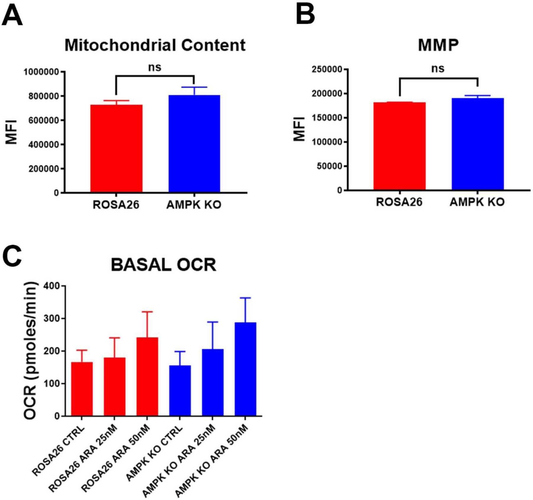
Figure 4. AMPK KO AML cells have intact mitochondria.

A) Mitotracker staining. AMPK KO and Rosa26 control cells were stained with the mitochondrial dye, mitotracker red. Shown are the mean values of the median florescence intensity (MFI) of three independent experiments. B) TMRM staining. AMPK KO and Rosa26 control cells were stained with the mitochondrial membrane dependent dye, TMRM. Shown are the mean values of the median florescence intensity (MFI) of three independent experiments. C) Basal oxygen consumption rates. AMPK KO and Rosa26 control cells were treated with the indicated amounts of cytarabine (ARA) for 16 hours and basal oxygen consumption rates (OCR) determined. Shown are means of three independent experiments each done in triplicate. There was no significant difference of OCR for any chemotherapy concentration.
