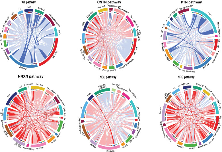
Fig. 5.
Chord diagrams plotting signaling strength differences between ASD and control PFC. The lines represent changes in L-R interaction strengths, with the statistically significantly different ones colored as intense red or blue, for increase or decrease in ASD, respectively. Light red or light blue for small changes not reaching statistical significance. Gray lines for no changes. Genes identified as differentially expressed in Velmeshev et al. [23] study were indicated in the corresponding cell type(s). The color bars in the inner circles indicates targeting cell types of the outgoing signaling while noncolor part for incoming signaling

