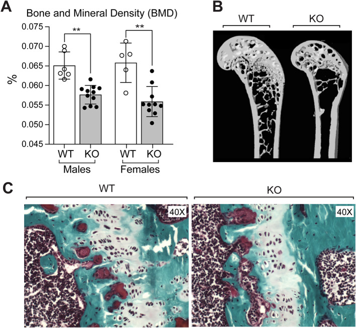
Fig. 2.

(A) Bone mineral density (BMD) assessed by dual‐energy X‐ray aborptiomtry (DXA) analysis in femurs of male and female mice at 7 months of age. (B) Reconstructed distal femur models generated from micro‐CT scans of 9‐month‐old WT and KO male mice. Data are presented as mean ± SD; statistical significance indicated on plots: **p < 0.01. (C) Histology of femoral growth plates of WT and KO mice. In each case, representative photomicrographs of the growth plate are shown stained by trichrome stain. Magnification ×40.
