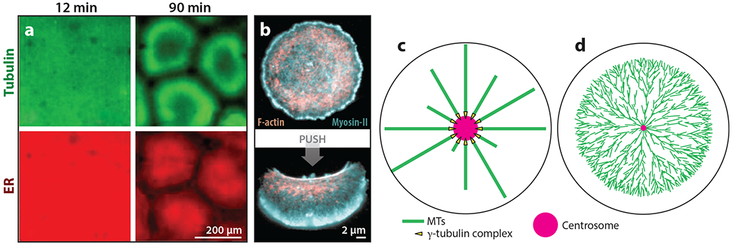
Figure 1.

Examples of self-organization and templating in cellular organization. (a) The self-organization of cellular units in interphase frog egg extract. Panel a adapted with permission from Cheng & Ferrell (2019). (b) An example of bistability in actomyosin-based cellular organization. Keratocyte fragments were stable in two organizational states, (top) circular/immobile and (bottom) polarized/migrating. Circular nonmotile fragments were converted into polarized motile fragments by pushing with a pipette. Panel b adapted from Verkhovsky et al. (1999). (c) The templating of MT organization by the centrosome in a small cell. (d) The self-organization of a MT aster in a frog egg, in which the aster grows as a traveling wave by MT-stimulated MT nucleation. The cell radius is ~600 μm and the average MT length is ~;15 μm. Panel d adapted from Ishihara et al. (2016). Abbreviations: ER, endoplasmic reticulum; MT, microtubule.
