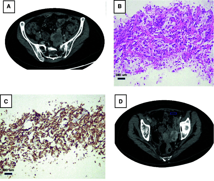
Figure 2.
Clinicopathological presentation of metastatic sarcomatoid urothelial carcinoma (UC). Computed tomography (CT) with contrast showing an enhancing left pelvic mass (arrow) near the pelvic sidewall and near the left internal iliac vessels (A,B). Biopsy from that mass showed sarcomatoid carcinoma related to patient's known bladder primary (C). PDL-1 (SP263 antibody) shows a diffuse membranous staining, and the combined positive score (CPS) was 100% (D).

