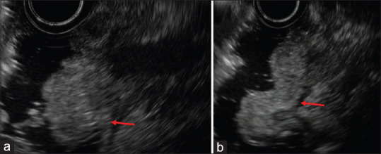
Figure 4.

Gel immersion-assisted EUS for ampullary lesions. (a and b) Representative images of ampullary lesions obtained by the gel immersion-assisted EUS are shown. The injected OS-1 gel remained in the duodenum for much longer than the water had due to its viscosity. Sharp EUS images with a clear lesion margin were obtained not only for the ampulla (a) but also for the anal side of the ampulla (b). The lesion was arising from the second layer of the duodenum
