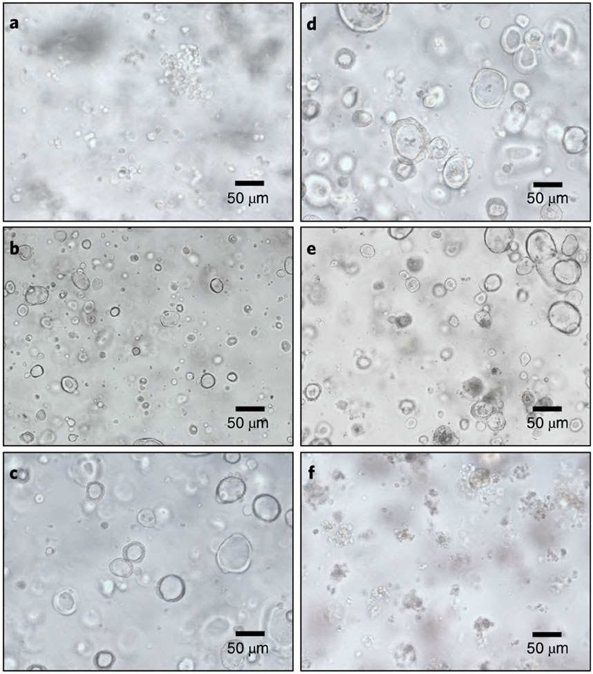
Fig. 3 ∣. HIE morphologies during transduction and puromycin selection.
a, Single-cell HIEs for lentiviral transduction at day 0. b, Expanded single-cell clones after 2 d of transduction. c,d, Expanded HIE clones 4 d after transduction, which are ready to be selected with puromycin. e, Transduced HIEs treated with puromycin for 4 d. f, Nontransduced HIEs treated with puromycin for 4 d with dying and dead cells. 20× objective lens.

