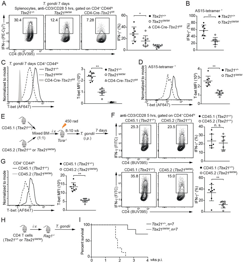
Figure 4. Defective Th1 cell differentiation in the Tbx215M/5M mice in response to T. gondii infection.
(A-D) Tbx21+/+ (n=8), Tbx215M/5M (n=8) and CD4-Cre-Tbx21fl/fl (n=8) mice were infected with T. gondii for 7 days. Splenocytes were stimulated with anti-CD3 and anti-CD28 antibodies for 5 hours in the presence of monensin. The percentage of IFN-γ-producing cells in the CD4+CD44hi population and in the T. gondii antigen AS15-specific CD4+ T cells was calculated (A and B). T-bet protein amounts were measured, and T-bet MFI was calculated (C and D). Mean ± SD.
(E-G) Experimental procedure of BM chimeras infected with T. gondii for 7 days (E). The percentage of IFN-γ-producing cells was calculated after anti-CD3 and anti-CD28 antibodies stimulation in the presence of monensin (F). T-bet protein amounts in the CD4+CD44hi T cells from CD45.1 Tbx21+/+ and CD45.2 Tbx215M/5M chimeras (n=8) were measured, and T-bet MFI was calculated (G). Mean ± SD.
(H and I) Negatively selected Tbx21+/+ or Tbx215M/5M CD4+ T cells were transferred into Rag1−/− recipients. The reconstituted mice (n=7 for each group) were infected with T. gondii, and then monitored for their survival.
Data are representative of two (A-I) independent experiments. See also Figure S3.

