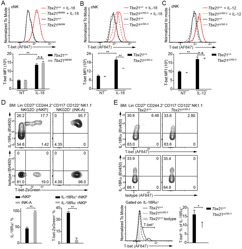
Figure 6. IL-18 up-regulates T-bet expression in cNK cells.
(A) Tbx21+/+ (n=3) and Tbx215M/5M (n=3) cNK cells were incubated with or without IL-18 for 3 days in the presence of IL-2. T-bet protein amounts were measured, and T-bet MFI was calculated. Mean ± SD.
(B and C) Tbx21+/+ (n=3) and Tbx21ΔCNS-3 (n=3) cNK cells were incubated with or without IL-18 (B) or with or without IL-12 (C) for 3 days in the presence of IL-2. T-bet protein amounts were measured, and T-bet MFI was calculated. Mean ± SD.
(D) The expression of IL-18Rα and T-bet reporter ZsGreen in the rNKPs and iNK-A cells from T-bet-ZsGreen reporter mouse BM was measured. The percentage of IL-18Rα+ cells in the rNKPs and iNK-A cells was calculated. The percentage of T-bet-ZsGreen+ cells in the IL-18Rα+ and IL-18Rα− rNKPs was calculated. Mean ± SD, n=3.
(E) The expression of T-bet in the bulk of rNKPs and iNK-A cells (Lin−CD27+CD244.2+CD117− CD122+NK1.1−) from Tbx21+/+ (n=3) and Tbx21ΔCNS-3 (n=3) mice was measured through intracellular staining. The percentage of T-bet+ cells in the IL-18Rα+ population was calculated. Mean ± SD.
Data are representative of two (A, C-E) or three (B) independent experiments. See also Figure S5 and S6.

