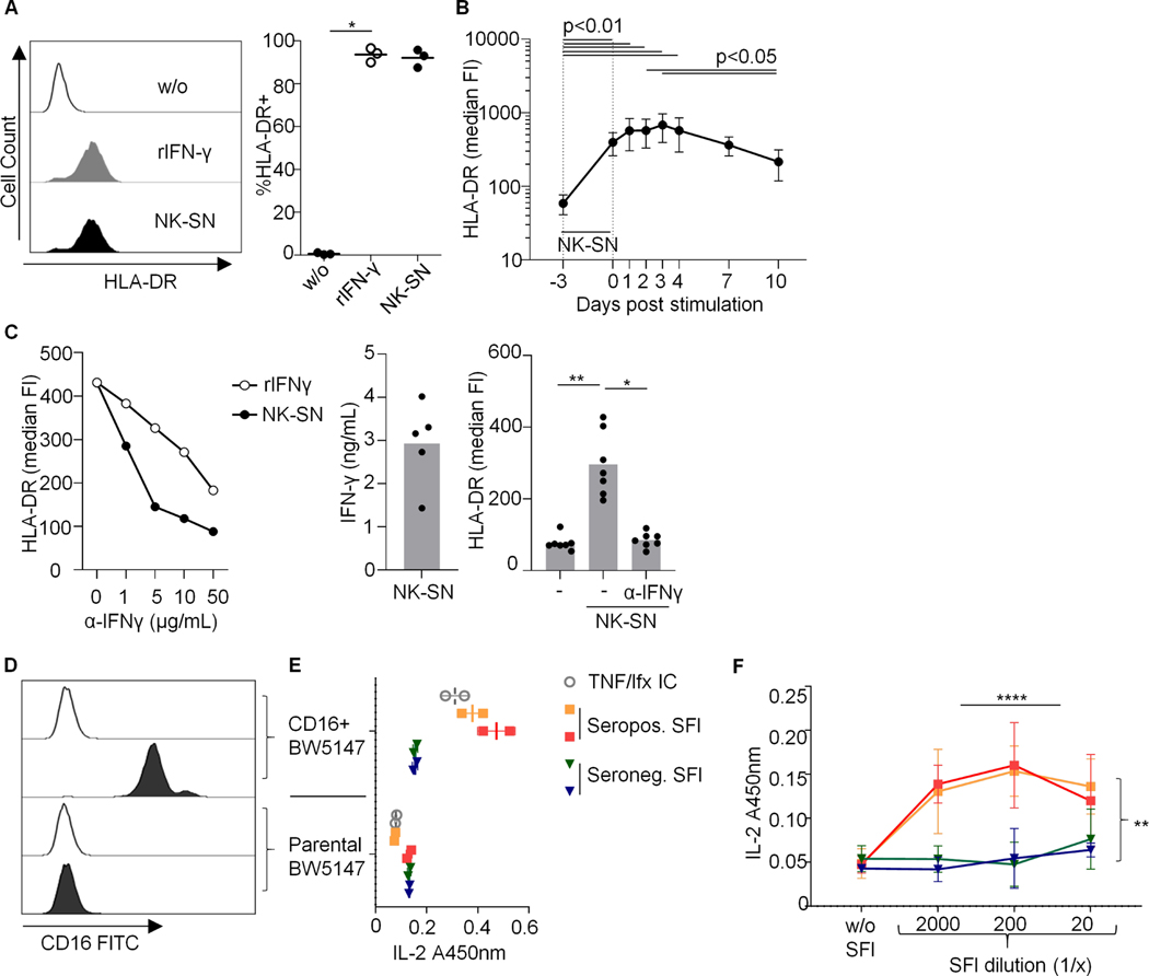
Fig. 1. (A-C) Induction of HLA-DR on fibroblasts by recombinant and NK cell-derived IFNɣ.
SFs were cultured over 3 days in medium (w/o) +/− recombinant (r)IFNɣ or pooled NK-SN and analyzed by flow cytometry. FI, fluorescence intensity. (A) Example histograms and percentages of HLA-DR+ RA-SFs, p=0.028. (B) HLA-DR kinetic (n=7 experiments; 3 OA-SFs; 4 RA-SFs); p<0.0001. (C) Effect of anti(α)-IFNɣ antibody on HLA-DR induction. Left: titration, n=1 experiment; middle: IFNɣ concentration in NK-SNs; right: 50μg/ml α-IFNɣ; n=7 experiments with three different RA-SFs, p=0.003. Statistics: Friedman test; significant post-tests are indicated. (D-F) Synovial fluid from seropositive RA activates CD16 (FcγRIIIA). Mouse-CD16+BW5147 reporter cells were cultured with synovial fluid (SFl) from two joints from an active seropositive RA patient or from each one joint from two patients with seronegative joint swelling (seronegative chronic polyarthritis and OA). (D) CD16 expression on BW5147 cells. Empty histograms: autofluorescence. (E) CD16 activation by SFl at a dilution of 1:1200, assessed by mouse-IL-2-ELISA (A450nm) (1 experiment in duplicates). TNFα + infliximab (TNF/Ifx) served as positive control. (F) CD16-activation by titrated SFl (n=3 technical replicates, each performed in triplicates). Three-way ANOVA confirmed significant effects of dilution and seropositivity (p<0.0001 and p=0.002). Bars: means +/− standard deviations.

