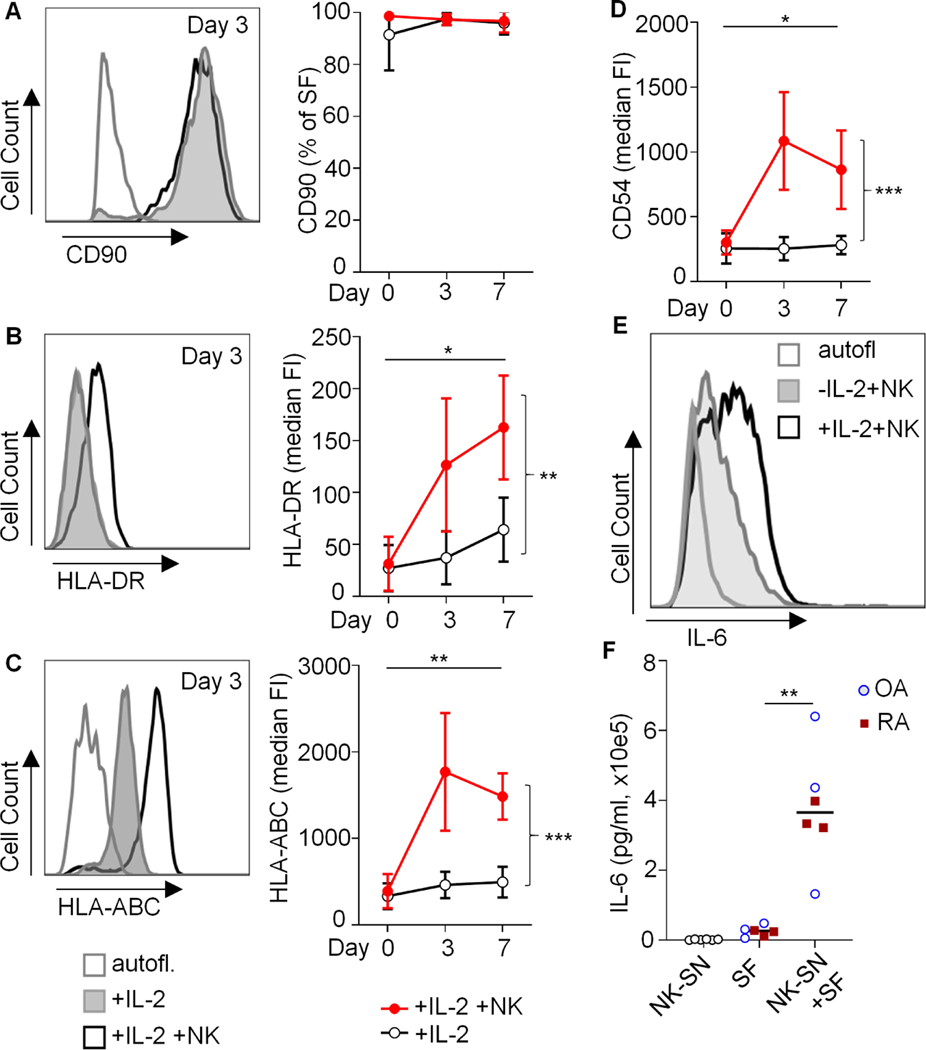
Fig. 4. Induction of HLA-DR and IL-6 in CD90+ synovial fibroblasts by activated NK cells in co-cultures.
(A-D) Synovial fibroblasts (SFs) from 3 OA patients were cultured in the absence or presence of IL-2-activated NK cells. The surface expressions of CD90, HLA-DR, HLA-ABC and CD54 (ICAM1) were analyzed by flow cytometry. Example histograms and means of median fluorescence intensities (FI) or means of percentages of positive cells and standard deviations are shown (n=3 identical experiments). Day 0: Untreated SFs. Statistical analysis: two-way ANOVA. *P<0.05, **P<0.01, ***P<0.001. Autofl.: autofluorescence. (E) Intracellular staining of IL-6 in OA-SF after direct co-culture with NK cells over three days. One of two similar experiments is shown. (F) For quantification of secreted IL-6, OA-SF and RA-SF (n=3 each) were cultured for 3 days +/− 20% pooled NK-SN. Supernatants were analyzed by ELISA. Significance was confirmed by Mann Whitney test (p=0.0022). In the same ELISA run, relevant amounts of IL-6 in NK-SN were excluded (n=6).

