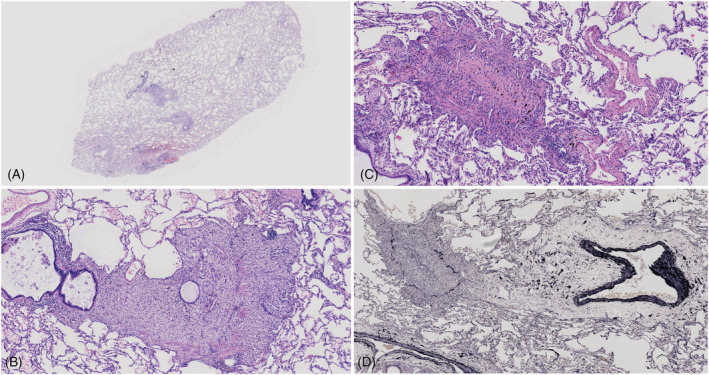
FIGURE 4.

Case 1: (A) Low‐magnification microscopic appearance of the left lower bronchiole. Lesions are exclusively limited to the area of the membranous bronchiole (original magnification ×40). (B) Granulation with plenty of foamy macrophages, with scattered giant cells causing stenosis of the bronchiolar lumen, whereas smooth muscle of the wall was partially destroyed. Epithelial sloughing and mucous retention are seen in the lumen (original magnification ×200). (C) Complete obliteration of the bronchial lumen due to submucosal concentric fibrosis (original magnification ×200). (D) There is total fibrous obliteration of the lumen (original magnification ×100). (A–C) Haematoxylin‐Eosin stain; (D) Elastic van Gieson stain
