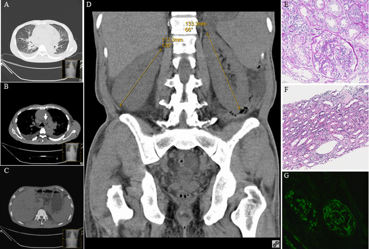
Fig 1. Computed tomography of the thorax and abdomen and histology of renal biopsy for Case 6.
(A) Bilateral diffuse interstitial infiltrates pleural effusion. (B) Bilateral pleural effusion and mediastinal lymphadenopathy (arrow). (C) Hepatosplenomegaly and ascites. (D) Symmetrically enlarged kidneys. (E) Diffuse intracapillary hyperplasia in the glomerulus with neutrophil infiltration in the capillary lumen, and mild proliferation of mesangial cells and stroma in focal segments of the glomerulus (PAS×400). (F) Focal renal interstitial fibrosis and edema with neutrophil, lymphocyte and plasmacyte infiltration (H&E×200). (G) Granular C3 deposition in the capillary wall and mesangial regions on immunofluorescent staining (×200).

