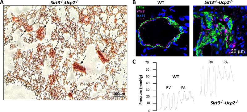
Figure 2. Plexogenic arteriopathy and severe PAH in mice lacking both Sirt3 and Ucp2.
(A) Representative histology (Hematoxylin-Eosin staining) of a lung from a Sirt3−/−;Ucp2−/− mouse shows numerous plexogenic lesions (back arrows). (B) Representative photomicrograph of confocal fluorescence immunohistochemistry of small pulmonary arteries from wild-type WT and Sirt3−/−;Ucp2−/− mice shows smooth muscle cells (green) and endothelial cells (magenta) in plexogenic lesions. (C) Representative tracings of RV and PA pressures from WT and Sirt3−/−;Ucp2−/− mice by close-chest right heart catheterization through the jugular vein shows that Sirt3−/−;Ucp2−/− mice have a significant increase in RV and PA pressure. SMA: smooth muscle actin, vWF: von Willebrand Factor (marking endothelial cells), DAPI (marking nuclei). (Images courtesy of Dr Gopinath Sutendra.)

