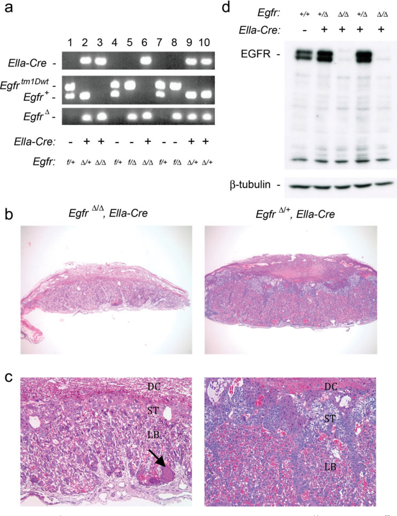
FIG. 3.
Analysis of EgfrΔ allele activity. (a) Genotypes of 12.5 dpc embryos from crosses between EgfrΔ/+, Ella-Cre, and Egfrf/f mice. (b) H&E staining of EgfrΔ/Δ, Ella-Cre placenta (left, ×30) with smaller size than wildtype placenta (right, ×30). (c) H&E staining of EgfrΔ/Δ, Ella-Cre placenta (left, ×100) showing reduced spongiotrophoblast layer and disorganized labyrinth layer with amorphous material (arrow) compared to wildtype placenta (right, ×100). (d) Western blot analysis showing loss of EGFR in EgfrΔ/Δ, Ella-Cre embryos at 10.5 dpc. DC, decidua; ST, spongiotrophoblast layer; LB, labyrinth layer.

