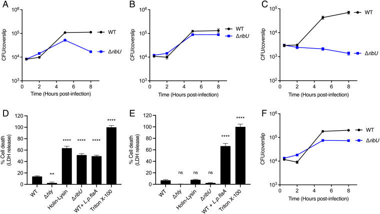
Fig. 2.
L. monocytogenes requires RibU to grow in riboflavin-starved macrophages. (A–C and F) Intracellular growth curves of L. monocytogenes strains in murine BMMs. BMMs were infected at a multiplicity of infection (MOI) of 0.1, and CFUs were enumerated at the indicated times. (A) Intracellular growth curves of indicated L. monocytogenes strains in WT BMMs. The data show the means and SEMs of four independent experiments. (B) Intracellular growth curves of indicated L. monocytogenes strains in WT BMMs incubated with cell culture media containing excess (10 µM) riboflavin during infection. The means and SEMs of three independent experiments are shown. (C) Intracellular growth curves of indicated flavin-starved L. monocytogenes strains in riboflavin-deficient WT BMMs. The data represent the means and SEMs of three independent experiments. (D and E) Cell death of WT (D) or AIM2 KO (E) BMMs infected with specified L. monocytogenes strains. LDH released to the cell culture media was used as an indicator of cell death. LDH release values were normalized to 1% Triton-X–treated cells which represent 100% lysis. BMMs were infected at an MOI of 4. The data show the means and SEMs of three technical replicates from at least two (D) and four (E) independent experiments. Statistical significance was determined using one-way ANOVA and Dunnett’s posttest using WT as the control. ****P < 0.0001; **P < 0.01; ns, not significant, P > 0.05. (F) Intracellular growth curves of indicated L. monocytogenes strains in AIM2 KO BMMs. The means and SEMs of five independent experiments are shown. WT + L.p.flaA, WT L. monocytogenes expressing Legionella pneumophila flagellin A under the control of the actA promoter (29). Holin-Lysin, WT L. monocytogenes expressing the bacteriophage proteins Holin and Lysin under the control of the actA promoter, leading to lysis of the bacteria in the cytosol of the host cell (28).

