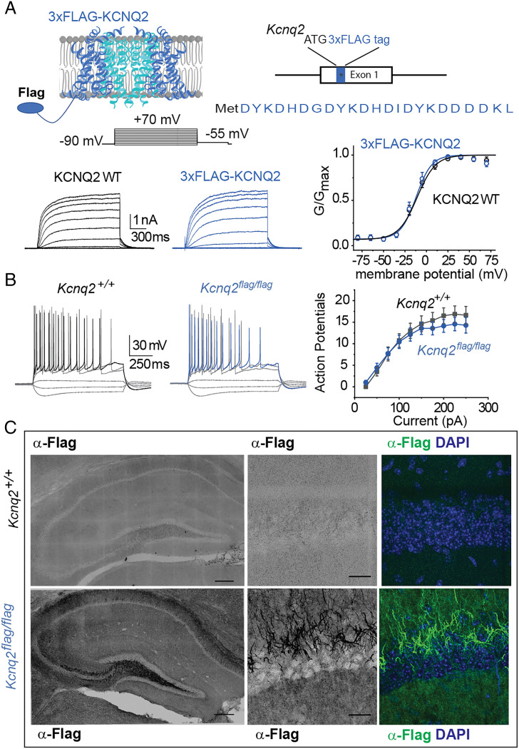
Fig. 3.
Development and characterization of Kcnq2 epitope-tagged mice. (A) Top, illustration showing the location of the epitope tag on the KCNQ2 channels and Kcnq2 gene. Bottom Left, representative recordings from cells expressing either wild-type KCNQ2 channels or 3XFLAG-tagged KCNQ2 channels. Bottom Right, summary graph showing that introduction of the 3XFLAG epitope does not change the properties of the KCNQ2 channels (wild-type V0.5= −13.7 ± 2.8 mV, n = 4; 3XFLAG-KCNQ2 V0.5 = −11.9 ± 2.6 mV, n = 5). Data are displayed as mean ± SEM (B) Left, representative recordings from either control CA1 pyramidal neurons or neurons expressing the 3XFLAG-tagged KCNQ2 channels. The membrane potential was held at −65 mV, maintained with small DC current injections. Right, summary graph showing that introduction of the 3XFLAG epitope does not change the firing properties of CA1 pyramidal neurons (Kcnq2+/+ n = 8, 2 mice; Kcnq2flag/flag n = 7; 2 mice). Data are displayed as mean ± SEM (C) Coronal sections showing localization of the 3XFLAG-tagged KCNQ2 channels in axons. Slices were stained with either DAPI (blue) or an anti-FLAG M2 antibody.

