
Figure 1.
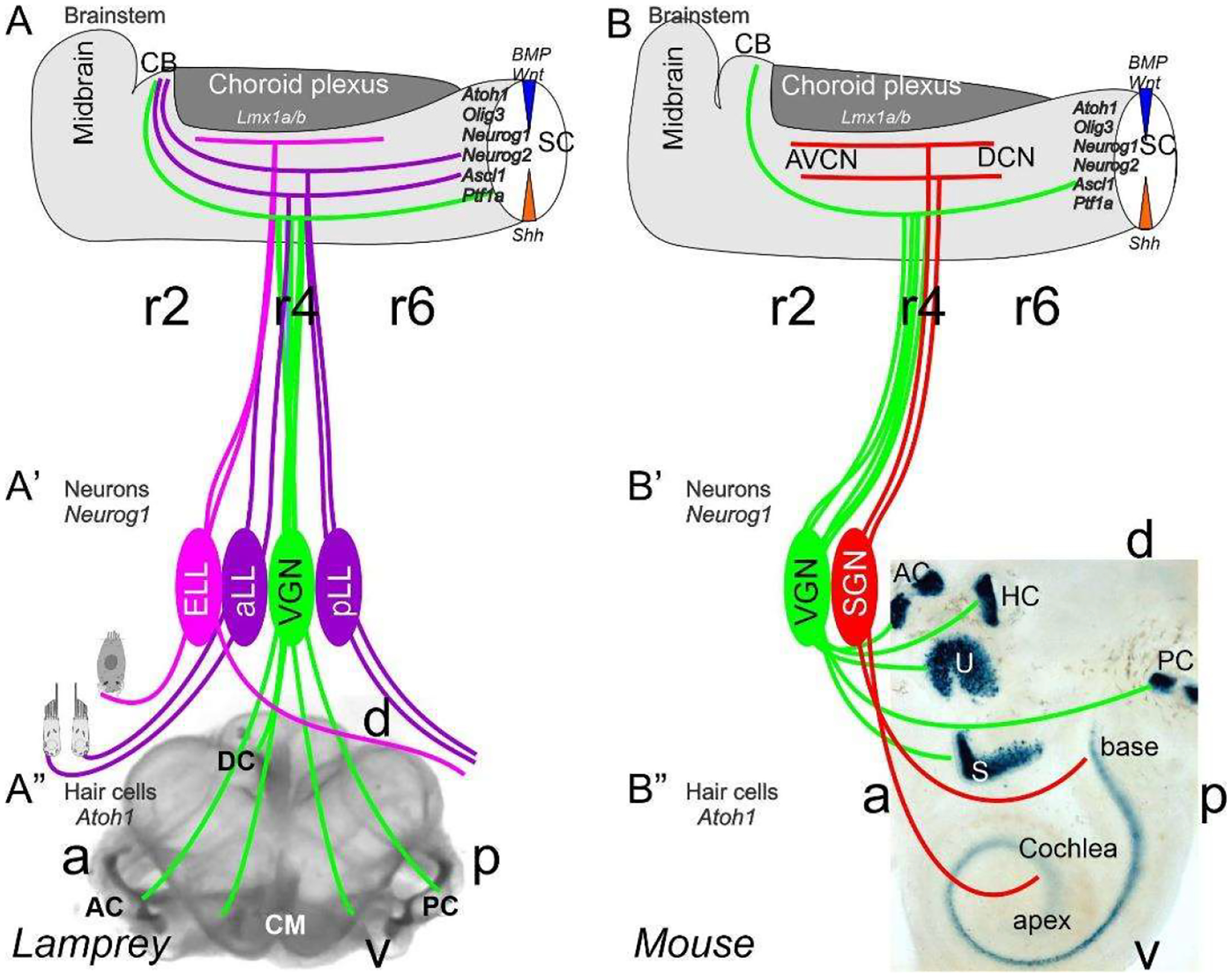
Inner ear, Lateral line and electroreception revealed. Neurons (Neurog1; A′) form vestibular ganglia (VGN) to reach out 4 hair cell organs in lampreys (A″). A separate lateral line (LL) and electroreceptor neurons (ELL) that innervate hair cells project more dorsal in lampreys. Central projection depends on Atoh1 to receive LL and ELL fibers, whereas several bHLH genes (Neurog1/2, Olig3, Ascla1, Ptf1a) receive all VGN (A). In the absence of ELL and LL development in amniotes, mammals develop separate spiral ganglion neurons (SGN; B′) that extend from the cochlea (B″) and end in a topological central projection that depends on Atoh1 (B). The formation of VGNs (Neurog1; B′) reach the 5 hair cells (B″) to extend the distribution of bHLH genes. Note that certain areas are lost or gained which enter central projections near r4. Images are shown by miR-183 ISH (A″) and Atoh1-LacZ (B″). AC, anterior crista; AVCN, anteroventral cochlear neurons; CB, cerebellum; aLL, pLL, anterior/posterior lateral line neurons; CM, common macula; DC, dorsal crista; DCN, dorsal cochlear neurons; HC, horizontal crista; PC, posterior crista; r2/4/6, rhombomeres; S, saccule; SC, spinal cord; U, utricle. Modified after [11,30,31].