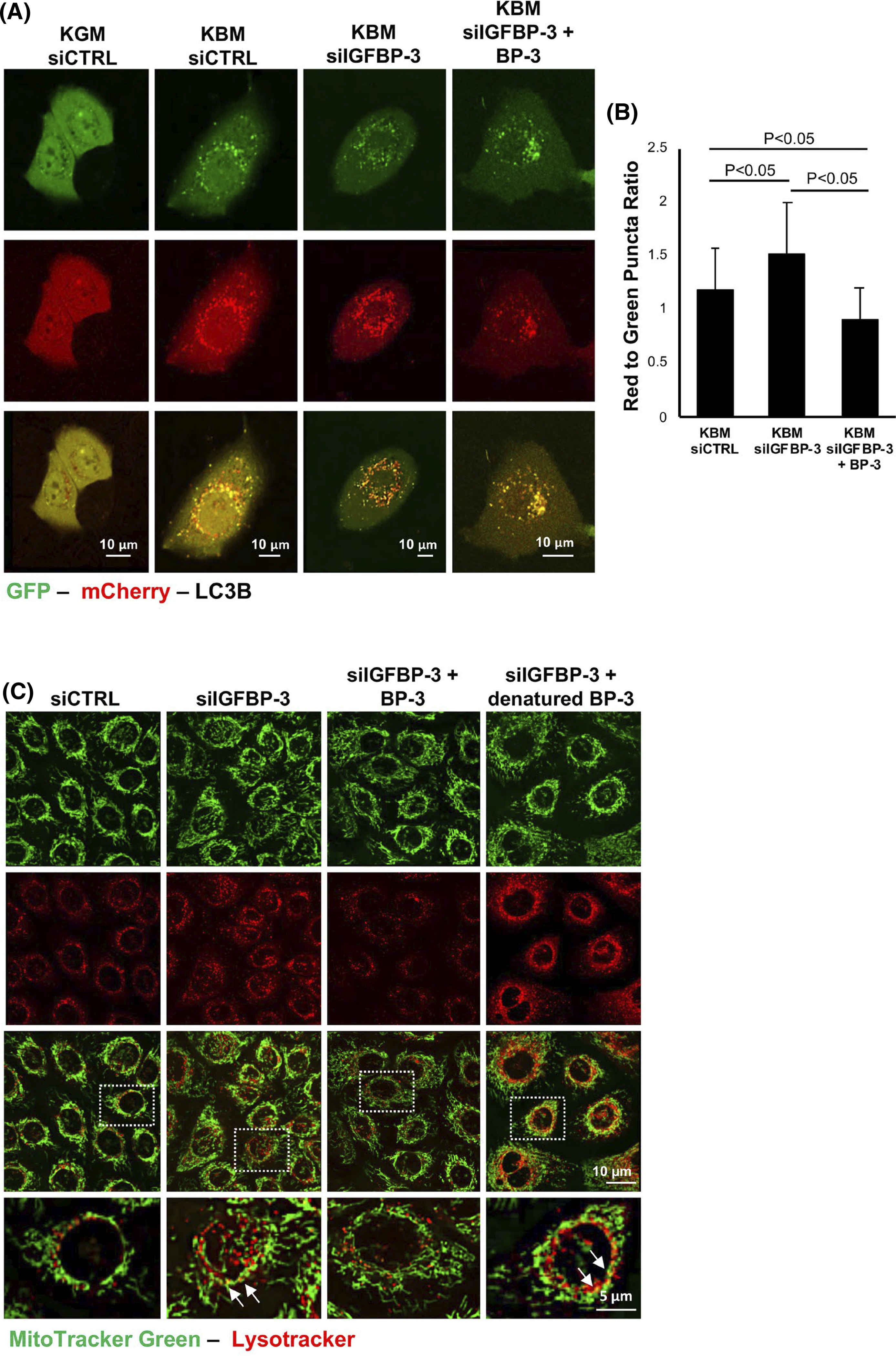
FIGURE 3.

IGFBP-3 inhibits autophagic flux in corneal epithelial cells. (A and B) hTCEpi cells were sequentially transfected with siRNA oligonucleotides targeting IGFBP-3 or a non-targeting control, followed by transfection with a GFP-mCherry-LC3 expression plasmid. Cells were then cultured in either KGM or KBM with or without 500 ng/ml rhIGFBP-3 for 24 h. (A) In KBM, there was an increase in the accumulation of both autophagosomes (yellow puncta) and autophagolysosomes (red puncta). Knockdown of IGFBP-3 increased the number of autophagolysosomes, whereas the addition of rhIGFPB-3 led to an accumulation of autophagosomes. Scale bar: 10 μm. (B) In IGFBP-3 knockdown cells, there was an increase in the ratio of red to green puncta compared to the siRNA control (p < .05). In contrast, co-treatment with rhIGFBP-3 showed a decrease in the ratio of red to green puncta compared to knockdown with IGFBP-3 and the KBM control (p < .05). (C) MitoTracker Green (green) and LysoTracker (red) colocalization in hTCEpi cells. Colocalization was rarely observed in the growth condition transfected with a control siRNA. There was a slight increase in colocalization in KBM. Knockdown of IGFBP-3 increased colocalization of both probes, indicating an increase in mitophagy. Treatment with rhIGFBP-3 decreased colocalization, suggesting an inhibition of mitophagy. Zoomed images corresponding to the white box in the merged image are shown in the bottom row. Arrows indicate areas of colocalization. Scale bar: 10 μm. Data presented as mean ± standard deviation from one representative experiment, N = 3. Fifteen cells per group were analyzed. KBM, keratinocyte basal media; KGM, keratinocyte growth media
