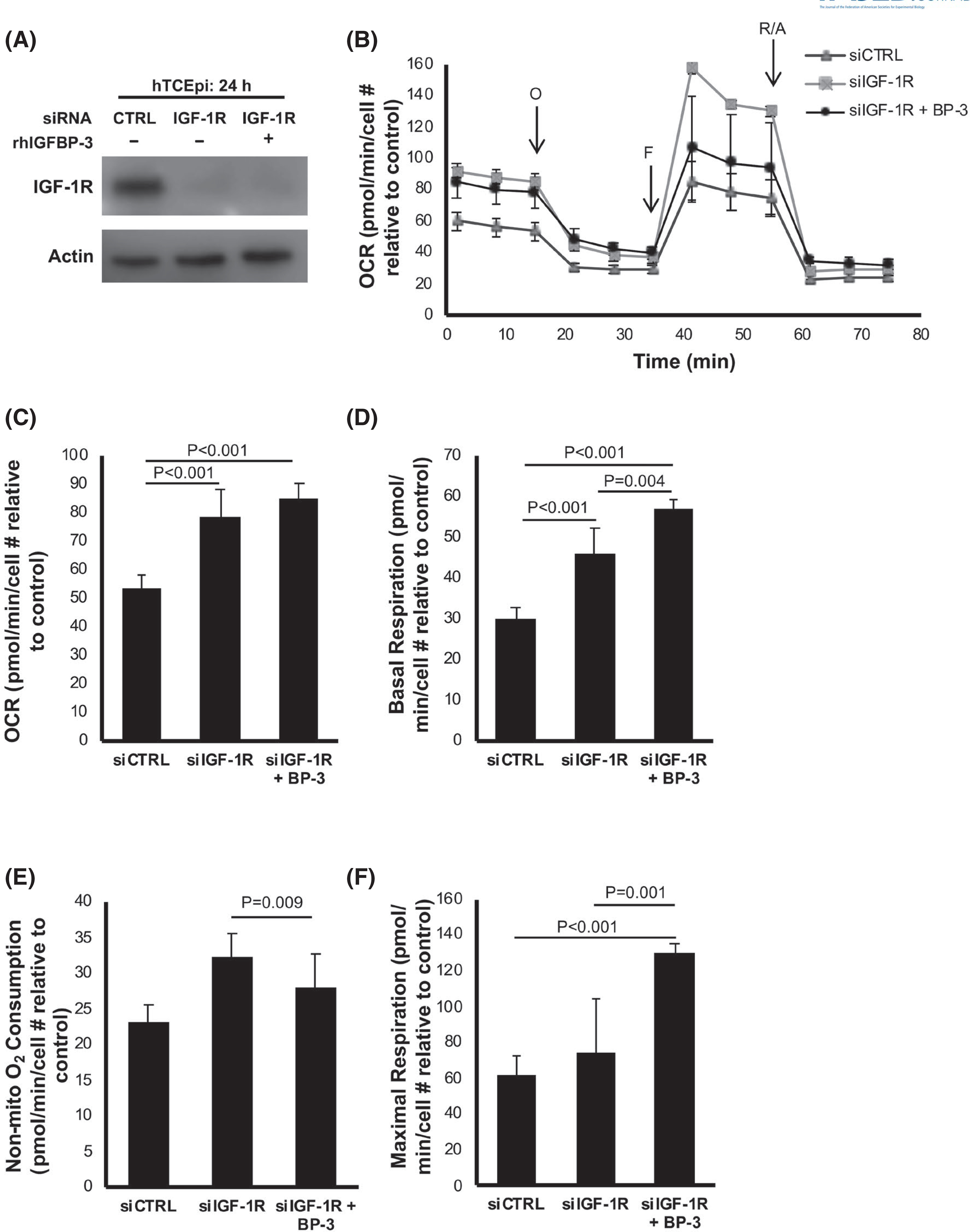
FIGURE 5.

IGF-1R regulates mitochondrial and non-mitochondrial oxygen consumption. hTCEpi cells were transfected with siRNA oligonucleotides targeting IGF-1R. Non-targeting oligonucleotides were used as a control. Cells were then cultured in KBM with or without 500 ng/ml rhIGFBP-3 for 24 h. (A) Immunoblotting was used to confirm knockdown of IGF-1R. ß-actin was used as a loading control. (B) OCR plotted as a function of time. The time points for addition of oligomycin (O), FCCP (F), and rotenone/antimycin A (R/A) are indicated. (C) OCR increased after IGF-1R knockdown (p < .001). OCR was unchanged in cells treated with rhIGFBP-3 compared to IGF1R knockdown. (D) IGF-1R knockdown increased basal OCR (p < .001), which was further increased by treatment with rhIGFBP-3 (p = .004). (E) Non-mitochondrial oxygen consumption was increased after IGF-1R knockdown (p = .009) but was unchanged with the addition of rhIGFBP-3 (F) Maximal respiration was not affected by IGF-1R knockdown, but was increased in cells co-treated with rhIGFBP-3 (p < .001 compared to control, p = .001 compared to IGF-1R knockdown). Data expressed as mean ± standard deviation from one representative experiment, N = 3. One-way ANOVA with Student–Newman–Keulspost hoc multiple comparison test. N = 3 repeated experiments. KBM, keratinocyte basal media; OCR, oxygen consumption rate
