Fig. 3.
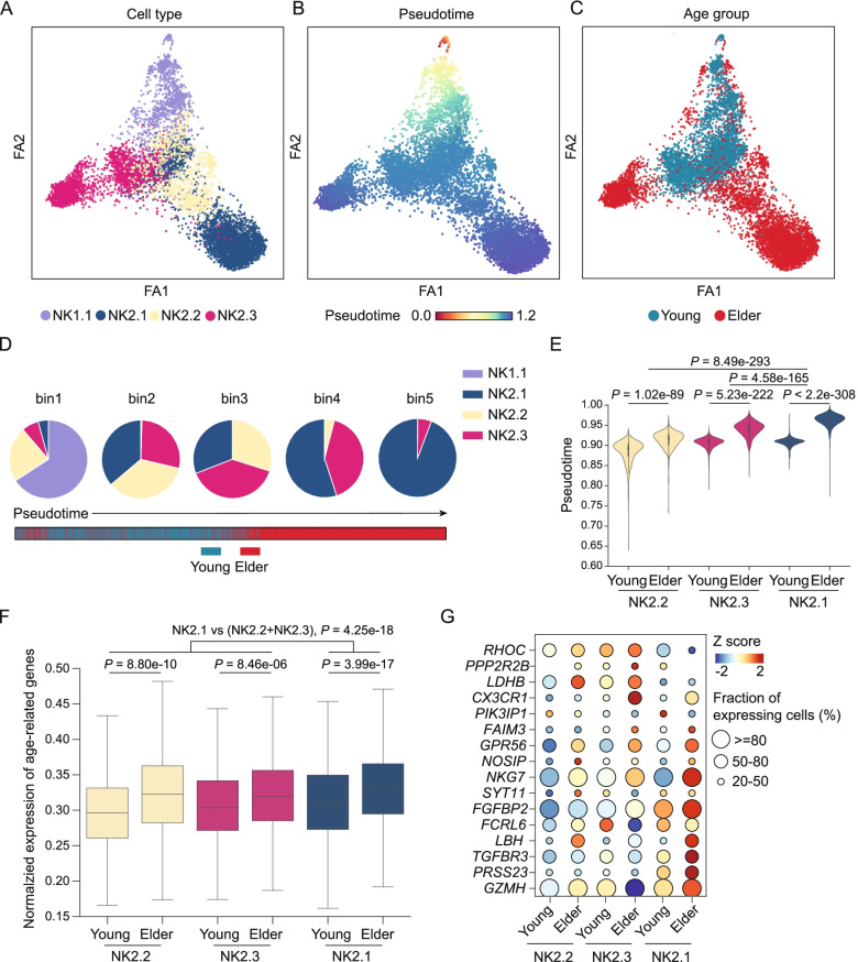
Pseudotime analysis reveals the distinct trajectories of memory-like NK cell differentiation during ageing. A–C Trajectories predicted using the PAGA algorithm for NK1.1, NK2.1, NK2.2, and NK2.3 cells from young and elderly individuals. Cells are coloured by NK cell subsets (A), by the pseudotime trajectory (B), and by age group (C). D Pie charts showing the composition of NK cell subsets in five bins; each bin was divided equally according to the pseudotime of the cells. E Violin plots of the cell pseudotime distribution for each NK cell subset from young and elderly individuals. P-values were estimated by Wilcoxon rank-sum test. F The box plot shows the expression of the top 50 age-associated genes (obtained from Peters et al. [31]) of NK2.1, NK2.2, and NK2.3 cells from young people and elderly people. P-values were obtained using the Wilcoxon rank-sum tests. G Dot plot representing the expression of age-related genes of NK2.1, NK2.2, and NK2.3 cells in the young and elderly groups. The plot shows genes expressed by at least 20% of the cells in the subset. The size of the dot represents the fraction of cells expressing the gene, and the colour of the dot represents the gene level as a z-score

