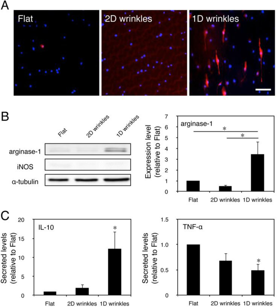
Figure 2. Surface topology regulates macrophage phenotype and cytokine secretion.
(A) Immunofluorescence images of arginase-1 on flat, 2D wrinkle, and 1D wrinkle surfaces. Scale bar = 50 μm. (B) Representative Western blot of arginase-1, iNOS, and α-tubulin on flat, 2D wrinkle, and 1D wrinkle surfaces (left) and quantification of arginase-1 expression of flat, 2D wrinkle, and 1D wrinkle surfaces (right). Mean ± SEM, astericks denotes p < 0.05; one-way ANOVA with Tukey’s posthoc test, n = 3. (C) Secretion of IL-10 (left) and TNF- α (right) by BMDM cultured on flat, 2D wrinkle, and 1D wrinkle surfaces for 24 hours. Mean ± SEM, astericks denotes p < 0.05 compared to flat surfaces; one-way ANOVA with Sidak’s posthoc test, n = 3.

