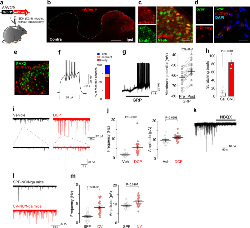
Fig. 1. Facilitation of glutamatergic synaptic inputs onto GRPR+ SDH neurons in mouse models of chronic itch.
a Schematic illustration of microinjection of AAV-GrprP-mCherry into the cervical SDH. b, c Visualization of mCherry+ cells (red) in the SDH (b), and immunolabeling of mCherry+ cells (red) by a pan-neuronal marker, NeuN (green) (c). d RNAscope in situ hybridization for Grpr mRNA (green) in mCherry+ cells (red) in the SDH. e Immunolabeling of mCherry+ cells (red) by an inhibitory neuronal marker, PAX2 (green). Scale bars, 200 μm (b), 50 μm (e), 20 μm (c, d). f Firing patterns of mCherry+ neurons in cervical spinal cord slices. Representative traces of delayed firing pattern evoked by injecting a depolarizing current, and the percentage of mCherry+ neurons displaying each pattern (n = 22 cells). g Representative response of GRP (200 nM) in mCherry+ neurons (displaying the delayed firing pattern), and the summary of the membrane potential of pre- and post-GRP application (n = 21 cells, paired t test). h Number of scratching behavior for 30 min after injection of clozapine-N-oxide (CNO, 10 mg/kg, i.p.) or saline in AAV-GrprP-hM3Dq mice (n = 4/group, unpaired t test). i, j Representative traces (i) and the average of frequency and amplitude (j) of sEPSCs in GRPR+ (mCherry+) SDH neurons of vehicle- and DCP-treated mice (vehicle, n = 11 cells; DCP, n = 14 cells; unpaired t test). k Representative traces of sEPSCs in GRPR+ (mCherry+) neurons of DCP-treated mice after application of NBQX, an antagonist for AMPARs. l, m Representative traces (l) and the frequency and amplitude (m) of sEPSC in SDH GRPR+ (mCherry+) neurons in SPF- and CV-NC/Nga mice (SPF, n = 16 cells; CV, n = 18 cells; unpaired t test). Values represent mean ± S.E.M. Source data are provided as a Source Data file.

