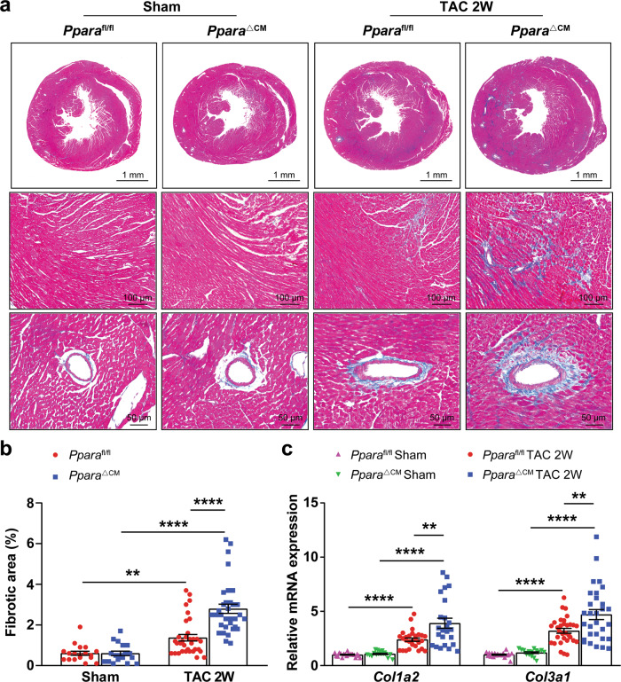
Fig. 2. Cardiomyocyte PPARα deficiency aggravated TAC-induced cardiac fibrosis.
Pparafl/fl and PparaΔCM mice were subjected to TAC or sham surgery, and analyses were performed 2 weeks later. a Representative images of Masson’s trichrome staining of heart sections. b Quantification of the fibrotic area (n = 18–33). c Expression of the fibrosis-related genes collagen type I alpha2 (Col1a2) and collagen type III alpha1 (Col3a1) in mouse hearts was detected by qPCR (n = 16–31). The data are expressed as the mean ± SEM. **P < 0.01 and ****P < 0.0001.

