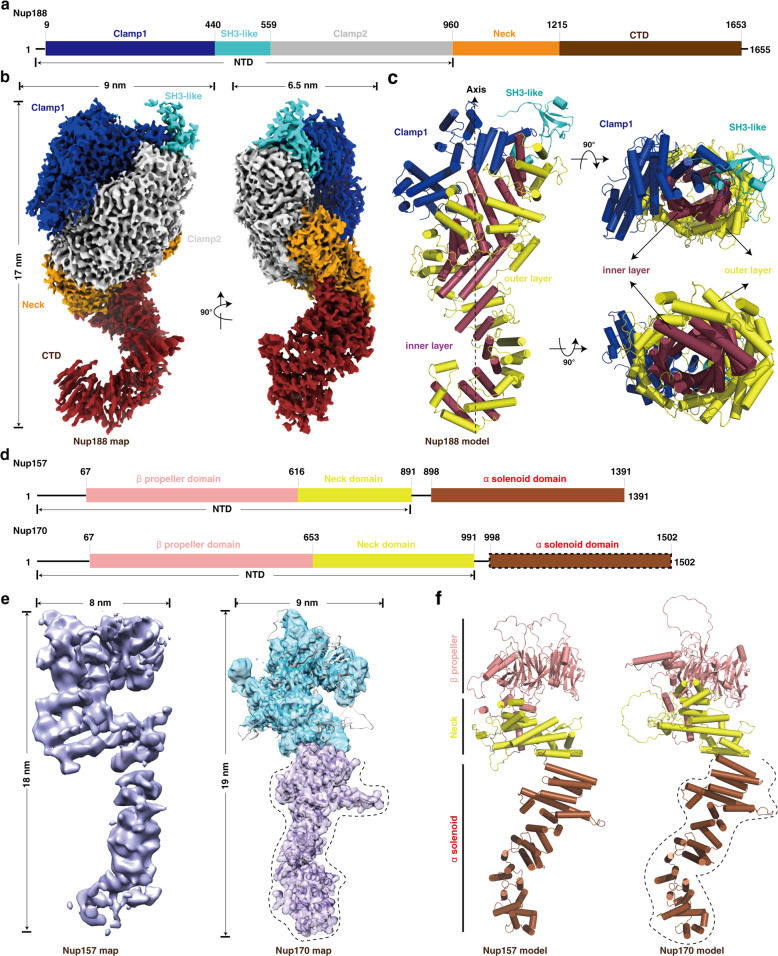
Fig. 2. Structures of the purified full-length Nup188, Nup157 and Nup170 proteins.
a Schematic representation of the domain structures of Nup188. Domains are color coded. b Final cryo-EM map and dimensions of Nup188. The contour level is 3.95 σ. Colors of domains are the same as in a. c Model of Nup188. Colors of Clamp1 and SH3-like domains are the same as in a, and the “S” domain is divided into two layers ― inner layer and outer layer, colored in raspberry and yellow, respectively. d Schematic representation of the domain structures of Nup157 and Nup170. Domains are color coded. The α-solenoid domain (boxed by dashed line) in Nup170 indicates missing regions in our map of Nup170. e, f Final cryo-EM maps (e) and models (f) of Nup157 and Nup170, respectively. The map of Nup170 is transparent and overlapped with its model. Colors of domains and dotted line marked regions are the same as in d. Cylinders represent α-helices. NTD, N-terminal domain; CTD, C-terminal domain.

