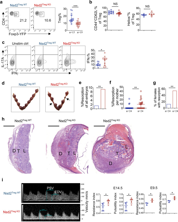
Fig. 2.
Nsd2 deficiency results in reduced Tregs at the maternal–fetal interface and in fetal resorption. a Representative flow cytometric analysis of YFP+ Treg cells in the decidua of Nsd2Treg WT and Nsd2Treg KO mice previously mated with male BALB/c mice on Days E13.5–E14.5. b Percentage of CD44+CD62Llow and Helios+ Tregs determined by flow cytometry in the decidua of Nsd2Treg WT and Nsd2Treg KO mice previously mated with male BALB/c mice on Days E13.5–E14.5. c Flow cytometric analysis of IFNγ and IL-17A expression in decidual CD4+ T cells. n = 10. d Representative image of resorption of allogeneic embryos in the uterus of Nsd2Treg WT and Nsd2Treg KO mice on Day E14.5. Arrows indicate resorptions. e Percentage of resorbed embryos in all Nsd2Treg WT and Nsd2Treg KO pregnancies resulting from matings with male BALB/c mice. Two-sided Fisher’s exact test was used to assess the significance. f Percentage of resorption observed in individual mothers with the indicated genotype. g Incidence of pregnancies with at least one resorption. Two-sided Fisher’s exact test was used to assess the significance. h Histopathological evaluation of placentas from female Nsd2Treg WT (left) and Nsd2Treg KO (right, two examples) mice mated with male BALB/c mice; low-magnification survey of representative H&E-stained sections of placenta. D decidua, T trophoblast, L labyrinths. Scale bars, 1000 mm. i Representative Doppler image of the uterine artery (left, E14.5), which was used to determine the uterine artery resistance and pulsatility indices (right) of Nsd2Treg WT and Nsd2Treg KO female mice on Days E14.5 and E9.5. *P ≤ 0.05; **P ≤ 0.01; ***P ≤ 0.001

