. 2019 Mar 18;9(16):8778–8881. doi: 10.1039/c8ra09577a

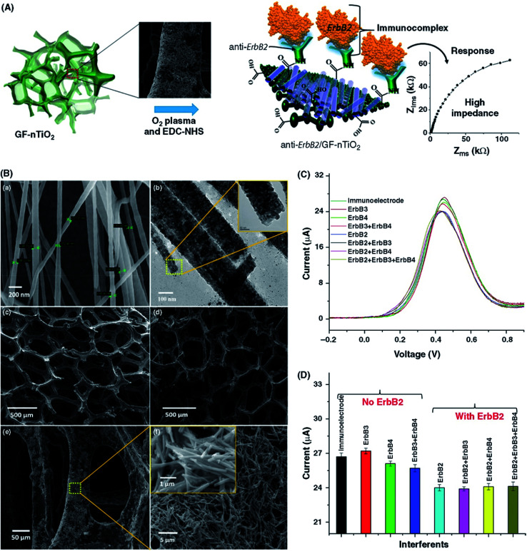
Fig. 26. (A) Functionalization of anti-ErbB2 molecules on the outer surfaces of GF and GF–nTiO2 electrodes. (B) FESEM (a) and TEM (b) images of carbon-doped nTiO2. The inset shows a magnified image of a single nTiO2. (c) SEM image of 3D GF. (d–f) SEM images of the GF–nTiO2 nanocomposite. (C) DPV curves of biosensor in the presence of ErbB3 and ErbB4 antigens. (D) Histogram displaying the peak current of the biosensor in the presence of different interfering bionanalytes. [Reprinted with permission from ref. 518, M. A. Ali, K. Mondal, Y. Jiao, S. Oren, Z. Xu, A. Sharma and L. Dong, Microfluidic Immuno-Biochip for Detection of Breast Cancer Biomarkers Using Hierarchical Composite of Porous Graphene and Titanium Dioxide Nanofibers, ACS Appl. Mater. Interfaces, 2016, 8, 20570–20582. Copyright© American Chemical Society.].

Fig. 26