. 2019 Mar 18;9(16):8778–8881. doi: 10.1039/c8ra09577a

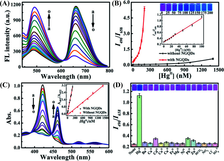
Fig. 37. (A) Fluorescence spectra of 5.0 μM 5,10,15,20-tetrakis(1-methyl-4-pyridinio)porphyrin tetra(p-toluenesulfonate) (TMPyP) after the addition of 20 μg L−1 N-GQDs, 40 μM MnII, and different concentrations of HgII ranging from 0 to 200 nM. (B) Plot of the fluorescence ratio (I490/I658) vs. HgII concentrations (0–200 nM) in the presence or absence (HgII concentrations of 0 to 1400 nM) of N-GQDs. Inset shows photographs corresponding to the HgII concentrations and the plot of (I490/I658) vs. HgII concentration (0–100 nM) in the presence of N-GQDs. (C) Absorption spectra of 5.0 μM TMPyP after the addition of 40 μM MnII, 20 μg L−1 N-GQDs, and different concentrations of HgII; inset shows plots of A462/A422vs. HgII concentration with and without N-GQDs. (D) The selectivity for HgII among different interfering analytes. The fluorescence quenching efficiency (I490/I658) of 5.0 μM TMPyP after the addition of 40 μM MnII, 100 nM HgII and 20 μg L−1 N-GQDs and other interfering ions, including BaII, CdII, CoII, CuII, FeII, NiII, PbII, ZnII, HCO3, Cl at 500 nM concentration; MgII, SO42−, NO3 at 100 μM concentration; and CaII at 200 μM concentration; inset shows photographs of the fluorescence changes under 365 nm UV light. [Reprinted with permission from ref. 705, D. Peng, L. Zhang, R.-P. Liang and J.-D. Qiu, Rapid Detection of Mercury Ions Based on Nitrogen-Doped Graphene Quantum Dots Accelerating Formation of Manganese Porphyrin, ACS Sens., 2018, 3, 1040–1047. Copyright© American Chemical Society.].

Fig. 37