. 2019 Mar 22;9(16):9331–9341. doi: 10.1039/c9ra00017h

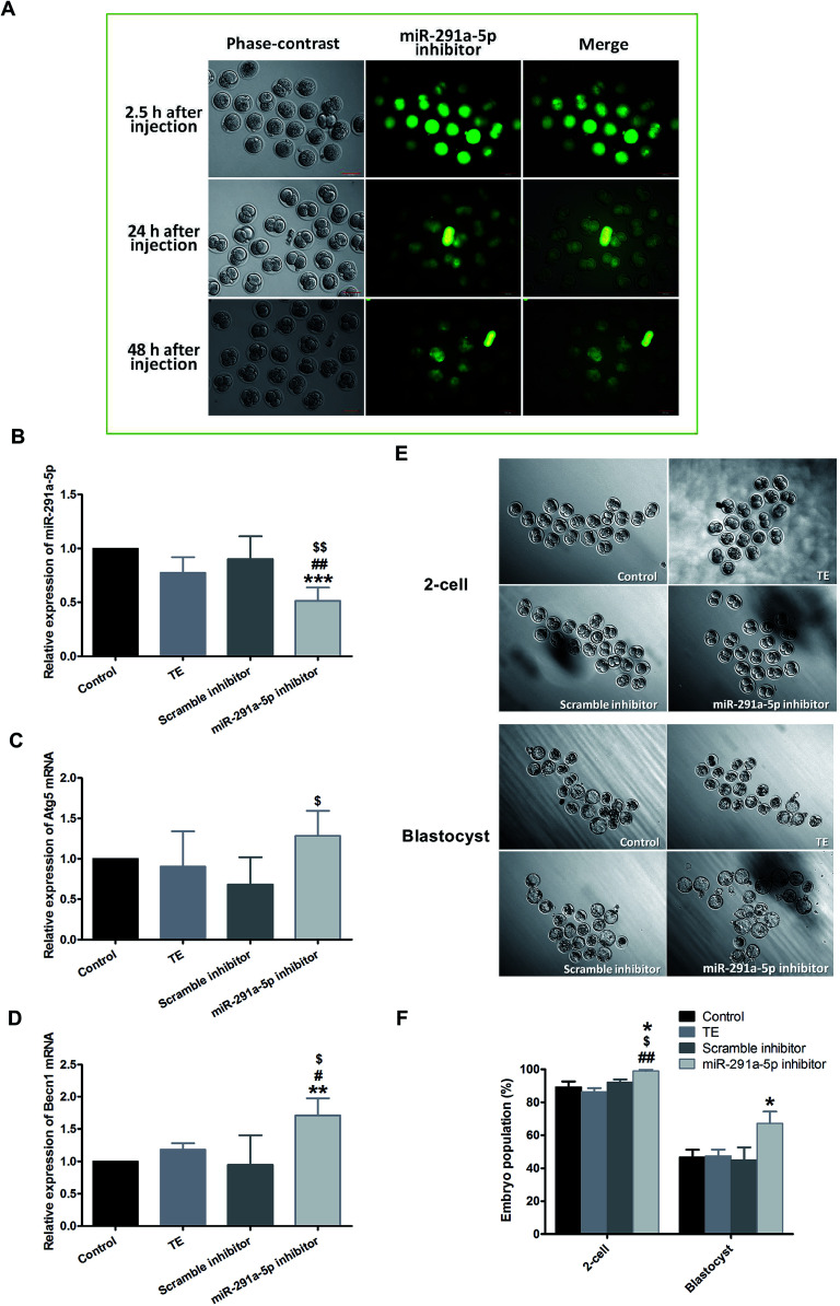
Fig. 4. (A) After microinjection of miR-291a-5p inhibitors, the expression of miR-291a-5p inhibitors in mouse embryos was observed using fluorescence microscopy. The mouse zygote was injected at 0.5 dpc with 5′ FAM-modified miR-291a-5p inhibitor molecules, which showed green fluorescence. 2.5 h, 24 h and 48 h after injection, green fluorescence was observed. Scale bar = 100 μm. After microinjection of the control, TE buffer, scramble inhibitor or miR-291a-5p inhibitor for 24 h, real-time quantitative PCR was performed to detect the expression of (B) miR-291a-5p, (C) Atg5 mRNA and (D) Becn1 mRNA in mouse embryos. $P < 0.05, $$P < 0.01 vs. scramble inhibitor group; #P < 0.05, ##P < 0.01 vs. TE group; **P < 0.01, ***P < 0.001 vs. control group. (E) After microinjection of control, TE buffer, scramble inhibitor or miR-291a-5p inhibitor for 24 h, the cleavage rates of mouse embryos at the 2-cell and blastocyst phases were observed under a microscope. (F) Analysis of mouse embryo development rate at 1.5 dpc (2-cell) and 4.5 dpc (blastocyst) after microinjection of various reagents, as above (n = 3, *P < 0.05 vs. control group, $P < 0.05 vs. scramble inhibitor group, ##P < 0.01 vs. TE group).

Fig. 4