
FIGURE 2.
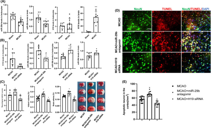
Changes in H19 and miR‐29b levels and subsequent effects on cerebral injury and neurological deficits in MCAO rats. (A) H19 and miR‐29b expression levels in leukocytes and ischemic brain tissues after MCAO injury (N = 9). (B) Quantitative RT‐PCR experiment to confirm the efficacy of H19 siRNA and miR‐29b antagomir transfection (N = 9). (C) Effects of intravenous injections of H19 siRNA and miR‐29b antagomir on cerebral injury and neurological function deficits at 24 h after stroke (N = 12). (D and E) Neuronal apoptosis in the ipsilateral cortex as detected by NeuN/TUNEL immunofluorescence double staining (N = 6). *p < 0.01, ***p < 0.001 vs. sham group. # p < 0.05, ### p < 0.001 vs. MCAO group. Scale bar = 20 μm