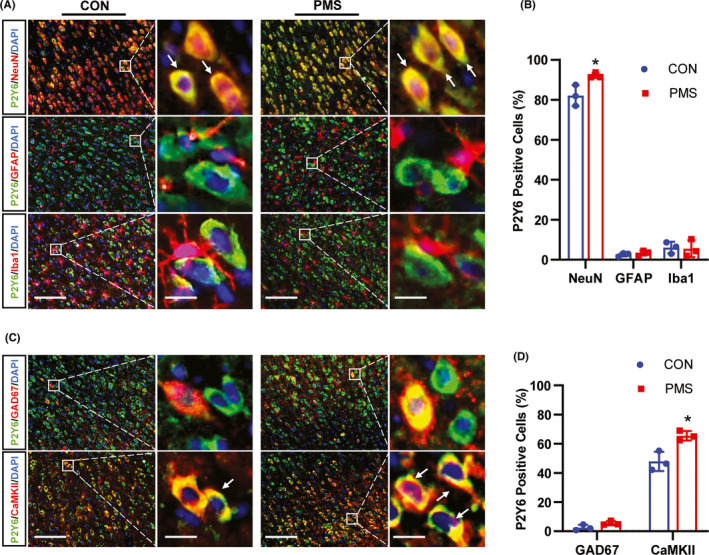
FIGURE 5.

PMS enhanced the percentage of P2Y6‐positive excitatory neurons in ACC. (A) Immunofluorescence study showed the co‐localization of P2Y6 with NeuN, GFAP, or Iba1 in ACC of CON and PMS rats (Bar = 50 μm). The enlarged pictures in the right panel showed the extent of co‐localization of P2Y6 (green) with NeuN (red), GFAP (red), or Iba1 (red) (Bar = 5 μm). The yellow staining indicated the co‐localization. (B) Data analysis showed that GRK6 was mainly localized in NeuN‐marked neurons, and a small amount in GFAP‐labeled astrocytes or Iba1‐dyed microglial in ACC. The ratio of GRK6‐positive cells over NeuN positive cells in the ACC region of PMS rats were significantly increased when compared with the CON rats (n = 3 for each group, *p < 0.05, two‐tailed two‐sample t‐test). (C) Immunofluorescence assay showed the co‐localization of P2Y6 with CaMKII or GAD67 (Bar = 50 μm). The enlarged pictures in the right panel showed the extent of co‐localization of P2Y6 with CaMKII or GAD67 (Bar = 5 μm). The yellow staining indicated the co‐localization. (D) Data analysis showed that P2Y6 was mainly present in CaMKII‐labeled excitatory neurons, and a small amount in GAD67‐labeled inhibitory neurons in ACC. The percent of P2Y6‐positive cells in CaMKII‐positive cells in the ACC region of PMS rats was significantly increased when compared with the CON rats (n = 3 for each group, *p < 0.05, two‐tailed two‐sample t‐test)
