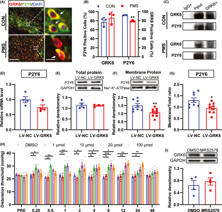
FIGURE 6.

GRK6 regulated the trafficking of P2Y6 in ACC. (A) Immunofluorescence assay showed the co‐localization of GRK6 and P2Y6 in ACC (Bar = 50 μm). (B) Quantity analysis showed that the ratio of GRK6‐positive cells over P2Y6‐positive cells in the ACC region of PMS rats was significantly decreased when compared with the CON rats. There was no significant difference of P2Y6‐positive cells over GRK6‐positive cells in the ACC region of PMS rats (n = 3 rats for each group, **p < 0.01, p > 0.05, two‐tailed two‐sample t‐test). (C) Co‐IP study confirmed the possible interaction of GRK6 and P2Y6 in the ACC region of both CON rats and PMS rats. (D‐E) LV‐GRK6 treatment did not affect the mRNA expression and the total protein expression of P2Y6 in ACC of PMS rats (n = 4 for each group, p > 0.05, two‐tailed two‐sample t‐test). (F) Compared with LV‐NC rats, the membrane protein expression of P2Y6 in LV‐GRK6 group was significantly reduced (n = 8 for LV‐NC group, n = 9 for LV‐GRK6 group, **p < 0.01, two‐tailed two‐sample t‐test). (G) The ratio of P2Y6 membrane protein expression to total protein expression in LV‐GRK6 group was significantly decreased (total protein level: n = 4 for each group; membrane protein level: n = 8 for LV‐NC group, n = 9 for LV‐GRK6 group, compared with LV‐NC rats, *p < 0.05, two‐tailed two‐sample t‐test). (H) P2Y6 selective inhibitor MRS2578 treatment significantly increased the CRD threshold of PMS rats (n = 6 for each group. *p < 0.05, **p < 0.01, ***p < 0.001, two‐way ANOVA). (I) The protein level of GRK6 was not altered in ACC of PMS rats treated with MRS2578 compared with DMSO‐treated group (n = 4 for each group, p > 0.05, two‐tailed two‐sample t‐test)
